Fluorescence Digital Image Gallery
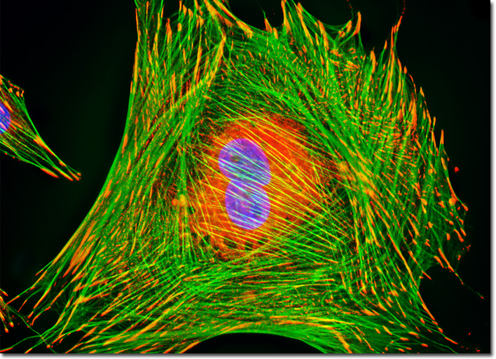
Indian Muntjac Deer Skin Fibroblast Cells
|
Vinculin is a protein associated with focal adhesion and adherens junctions, which are membrane-associated complexes that serve as nucleation sites for actin filaments and as crosslinkers between the external medium, plasma membrane, and actin cytoskeleton. Recently, vinculin has become a central focus of a significant amount of scientific research, resulting in the development of a better understanding of its role in cellular adhesion, motility, and intercellular communication, which appears to vary depending upon alterations in the three-dimensional structure of the protein. Studies have shown, for instance, that by alternating between an active and inactive form, vinculin can regulate the mobility of cells. Indeed, vinculin may facilitate cell movement for wound healing and embryonic tissue development, but is more interesting to some researchers for its possible involvement in the metastasis of cancerous tumors. The culture of Indian Muntjac deer skin fibroblast cells featured in the digital image above was immunofluorescently labeled with primary anti-vinculin mouse monoclonal antibodies followed by goat anti-mouse Fab heavy and light chain fragments conjugated to Cy3 (red fluorescence emission). In addition, the specimen was simultaneously stained for DNA with the ultraviolet-absorbing probe Hoechst 33258, and for the cytoskeletal filamentous actin network with Alexa Fluor 488 conjugated to phalloidin. Images were recorded in grayscale with a QImaging Retiga Fast-EXi camera system coupled to an Olympus BX-51 microscope equipped with bandpass emission fluorescence filter optical blocks provided by Omega Optical. During the processing stage, individual image channels were pseudocolored with RGB values corresponding to each of the fluorophore emission spectral profiles. |
© 1995-2025 by Michael W. Davidson and The Florida State University. All Rights Reserved. No images, graphics, software, scripts, or applets may be reproduced or used in any manner without permission from the copyright holders. Use of this website means you agree to all of the Legal Terms and Conditions set forth by the owners.
This website is maintained by our
|