Fluorescence Digital Image Gallery
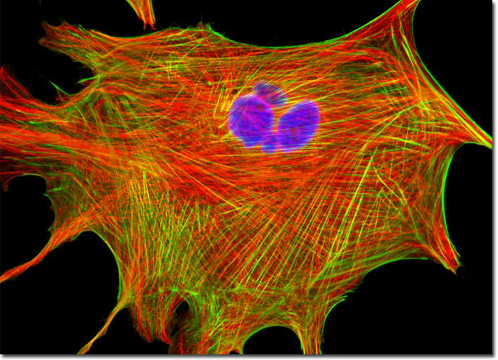
Indian Muntjac Deer Skin Fibroblast Cells
|
DAPI (4',6-diamidino-2-phenylindole) is a commonly utilized nucleic acid and chromosome counterstain that was originally synthesized as a potential anti-trypanosomal agent. Though impermeant at low concentrations (nanomolar), DAPI will penetrate the plasma membrane of live cells at higher concentrations. The blue fluorescent stain is highly DNA specific, and little or no cytoplasmic labeling usually occurs at concentrations less than 100 nanograms per milliliter. Indeed, studies indicate that DAPI binds preferentially to adenosine and thymidine (A-T) base pair regions in DNA, and in the bound state the fluorescence quantum yield is significantly enhanced. DAPI, which has a relatively large Stokes shift, is excited by ultraviolet light with a maximum absorption wavelength peak ranging between 350 to 360 nanometers and has a corresponding emission maximum of approximately 460 nanometers. The digital image presented above features a resident cell from a culture of Indian Muntjac fibroblasts that was labeled for DNA with DAPI, and for the cytoskeletal filamentous actin network with Alexa Fluor 488 conjugated to phalloidin. In addition, the culture was immunofluorescently labeled with primary anti-tubulin mouse monoclonal antibodies followed by goat anti-mouse Fab fragments conjugated to Rhodamine Red-X. Images were recorded in grayscale with a QImaging Retiga Fast-EXi camera system coupled to an Olympus BX-51 microscope equipped with bandpass emission fluorescence filter optical blocks provided by Omega Optical. During the processing stage, individual image channels were pseudocolored with RGB values corresponding to each of the fluorophore emission spectral profiles. |
© 1995-2025 by Michael W. Davidson and The Florida State University. All Rights Reserved. No images, graphics, software, scripts, or applets may be reproduced or used in any manner without permission from the copyright holders. Use of this website means you agree to all of the Legal Terms and Conditions set forth by the owners.
This website is maintained by our
|