Fluorescence Digital Image Gallery
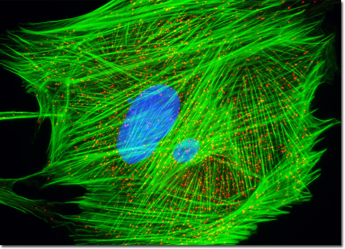
Indian Muntjac Deer Skin Fibroblast Cells
|
Discovered in 1965 by Christian de Duve, peroxisomes are multifunctional organelles present in almost all eukaryotic cells. These organelles were named for the hydrogen peroxide they produce as an intermediate in the process of normal cellular metabolism. Through the use of the enzyme catalase, peroxisomes are also responsible for detoxifying cellular metabolic side products by converting the hydrogen peroxide they generate into water and free oxygen molecules. The prolific single-membrane organelles are involved in a number of other cellular activities as well, including the degradation of lipids and the synthesis of various biochemicals. Peroxisomal membrane protein 70 (PMP 70) is a key membrane component of peroxisomes that can be readily utilized as a target to image these organelles using immunofluorescence microscopy techniques. The peroxisome organelles present in the Indian Muntjac fibroblast cell culture illustrated above were immunofluorescently labeled with Rhodamine Red-X conjugated to antibodies directed against peroxisomal membrane protein 70 (PMP 70). Alexa Fluor 488 conjugated to phalloidin and Hoechst 33258 were simultaneously used to counterstain the culture, targeting F-actin and DNA, respectively. Images were recorded in grayscale with a QImaging Retiga Fast-EXi camera system coupled to an Olympus BX-51 microscope equipped with bandpass emission fluorescence filter optical blocks provided by Omega Optical. During the processing stage, individual image channels were pseudocolored with RGB values corresponding to each of the fluorophore emission spectral profiles. |
© 1995-2025 by Michael W. Davidson and The Florida State University. All Rights Reserved. No images, graphics, software, scripts, or applets may be reproduced or used in any manner without permission from the copyright holders. Use of this website means you agree to all of the Legal Terms and Conditions set forth by the owners.
This website is maintained by our
|