Fluorescence Digital Image Gallery
Indian Muntjac Deer Skin Fibroblast Cells
|
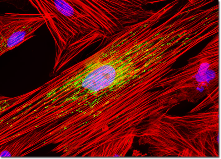
Localization of specific peptides, proteins, and macromolecular complexes to the mitochondria in mammalian cells is typically achieved through the utilization of peptide signals that mediate transport to the organelles. Recombinant plasmids have been produced that contain a fusion protein consisting of enhanced yellow fluorescent protein (EYFP), a yellow-green variant of the popular Aequorea victoria green fluorescent protein (GFP), coupled to the mitochondrial targeting nucleotide sequence from subunit VIII of human cytochrome C oxidase. Once transcription and translation of the plasmid occurs in transfected mammalian hosts, the mitochondrial localization signal transports and distributes the fluorescent protein chimera throughout the mitochondrial network of cultured cells, enabling the visualization of tubular mitochondria using fluorescence microscopy. The fluorescence absorption maximum of EYFP resides at approximately 513 nanometers and the corresponding emission maximum occurs in the region of 527 nanometers. The culture of Indian Muntjac deer skin cells presented in the digital image above was transfected with a pEYFP-Mitochondria (enhanced yellow fluorescent protein) chimeric plasmid subcellular localization vector. After fixation and permeabilization, the cells were labeled with Alexa Fluor 568 conjugated to phalloidin and DAPI, which target the F-actin cytoskeletal network and DNA in the cell nucleus, respectively. Images were recorded in grayscale with a QImaging Retiga Fast-EXi camera system coupled to an Olympus BX-51 microscope equipped with bandpass emission fluorescence filter optical blocks provided by Omega Optical. During the processing stage, individual image channels were pseudocolored with RGB values corresponding to each of the fluorophore emission spectral profiles. |
© 1995-2025 by Michael W. Davidson and The Florida State University. All Rights Reserved. No images, graphics, software, scripts, or applets may be reproduced or used in any manner without permission from the copyright holders. Use of this website means you agree to all of the Legal Terms and Conditions set forth by the owners.
This website is maintained by our
|