Fluorescence Digital Image Gallery
Indian Muntjac Deer Skin Fibroblast Cells
|
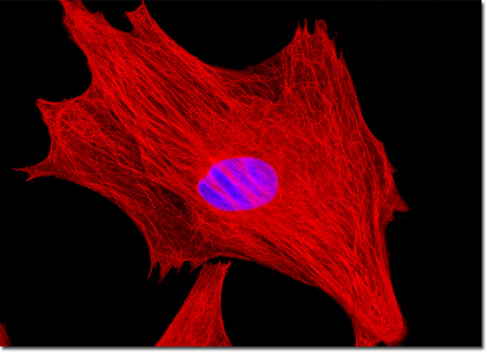
Hoechst 33258 is a bisbenzimide heterocyclic fluorescent probe that is permeant to the membranes of both living and dead cells. The dye is, however, reportedly less permeable to membranes than some other commonly utilized Hoechst probes. Nevertheless, Hoechst 33258 is a very popular nucleic acid stain, which fluoresces bright blue when bound to the minor groove of DNA, though it may also bind to RNA, albeit with significantly lower binding constant and quantum yield. Hoechst 33258 is relatively soluble in water and exhibits pH-dependent spectra when not bound to nucleic acids, with a considerably greater fluorescence quantum yield at pH 5 than at pH 8. When the dye is bound to DNA, it exhibits absorption and emission maxima of approximately 346 and 460 nanometers, respectively. The expansive cellular microtubule network present in a culture of Indian Muntjac fibroblasts (illustrated above) was immunofluorescently labeled with primary anti-tubulin mouse monoclonal antibodies followed by goat anti-mouse Fab fragments conjugated to Rhodamine Red-X. The culture was also counterstained for DNA in the cell nucleus with Hoechst 33258. Images were recorded in grayscale with a QImaging Retiga Fast-EXi camera system coupled to an Olympus BX-51 microscope equipped with bandpass emission fluorescence filter optical blocks provided by Omega Optical. During the processing stage, individual image channels were pseudocolored with RGB values corresponding to each of the fluorophore emission spectral profiles. |
© 1995-2025 by Michael W. Davidson and The Florida State University. All Rights Reserved. No images, graphics, software, scripts, or applets may be reproduced or used in any manner without permission from the copyright holders. Use of this website means you agree to all of the Legal Terms and Conditions set forth by the owners.
This website is maintained by our
|