Fluorescence Digital Image Gallery
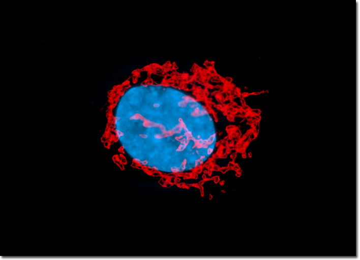
Human Fetal Lung Fibroblast Cells (MRC-5)
|
The Golgi apparatus, which is also known as the Golgi complex, was one of the first organelles ever identified with a light microscope. Indeed, the relatively large size and regular configuration of the Golgi apparatus makes it a very prominent cellular feature. Existing as a group of flattened sac-like structures known as cisternae, each of which is surrounded by a membrane, the appearance of the Golgi complex has often been compared to that of a stack of pancakes or pita bread. The number of cisternae in a Golgi stack varies and multiple stacks may be found in the same cell. In some types of cells, several Golgi stacks are connected to one another via tubular linkages, resulting in a sizable complex typically located near the centrosome adjacent to the nucleus. Vesicles concentrated in the area surrounding the Golgi apparatus are generally in the process of transferring material between the organelle and other cellular structures. In order to examine structural features of the Golgi complex and nucleus at relatively high magnification, a log-phase culture of MRC-5 cells (illustrated above) was fixed, permeabilized, blocked with normal goat serum, and then treated with rabbit anti-giantin (Golgi protein) primary antibodies followed by goat anti-rabbit secondary antibodies (IgG) conjugated to Alexa Fluor 568. The nuclei were counterstained with Hoechst 33258. Images were recorded in grayscale with a QImaging Retiga Fast-EXi camera system coupled to an Olympus BX-51 microscope equipped with bandpass emission fluorescence filter optical blocks provided by Omega Optical. During the processing stage, individual image channels were pseudocolored with RGB values corresponding to each of the fluorophore emission spectral profiles. |
© 1995-2025 by Michael W. Davidson and The Florida State University. All Rights Reserved. No images, graphics, software, scripts, or applets may be reproduced or used in any manner without permission from the copyright holders. Use of this website means you agree to all of the Legal Terms and Conditions set forth by the owners.
This website is maintained by our
|