Fluorescence Digital Image Gallery
Pig Kidney Epithelial Cells (LLC-PK1)
|
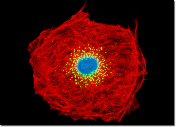
Antibodies are generated by immunizing susceptible hosts, such as rabbits, mice, goats, chickens, and other animals, with a specific antigen. If the antigen is derived from tissue, it must be exhaustively purified, but many antigens are now synthetic portions of a larger protein or carbohydrate. If a large protein is used as the antigen, such as a multi-subunit globular protein, the antibodies produced by the donor animal are not likely to be pure, but will usually be directed towards several portions (in effect, the subunits) of the large conglomerate. These antibodies are referred to as being polyclonal, and usually contain antibodies that react to carrier proteins and other native antibodies that may react to other tissue components. In this case, immunological reactions are usually not specific, targeting the desired antigen-antibody reaction, and are often complicated by high levels of background noise. Many epithelial cell cultures are good candidates for immunofluorescence labeling with cytokeratin. The LLC-PK1 culture presented above was treated with mouse anti-cytokeratin (pan) primary antibodies followed by goat anti-mouse secondaries conjugated to Texas Red. Simultaneously, the Golgi complex was visualized with rabbit anti-giantin primaries and goat anti-rabbit secondaries conjugated to Oregon Green 488. Nuclei were counterstained with Hoechst 33342. Images were recorded in grayscale with a QImaging Retiga Fast-EXi camera system coupled to an Olympus BX-51 microscope equipped with bandpass emission fluorescence filter optical blocks provided by Omega Optical. During the processing stage, individual image channels were pseudocolored with RGB values corresponding to each of the fluorophore emission spectral profiles. |
© 1995-2025 by Michael W. Davidson and The Florida State University. All Rights Reserved. No images, graphics, software, scripts, or applets may be reproduced or used in any manner without permission from the copyright holders. Use of this website means you agree to all of the Legal Terms and Conditions set forth by the owners.
This website is maintained by our
|