Fluorescence Digital Image Gallery
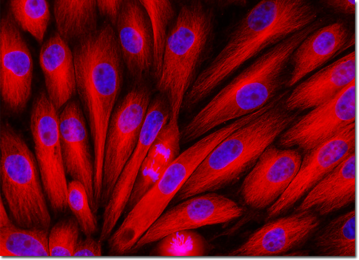
Pig Kidney Epithelial Cells (LLC-PK1)
|
Tubulin is a globular protein that is the chief structural component of microtubules. Though generally shaped into hollow fibers, microtubules, which act as a skeletal system for living cells, are capable of shifts in formation that enable mitosis and the regulation of intracellular transport. Tubulin, which is characteristically flexible, is responsible for this unusual ability of microtubules. Scientific interest in tubulin exploded in recent years due to the discovery that taxol, a substance originally derived from the bark of the Pacific yew tree, is able to battle many forms of cancer by binding to tubulin, essentially causing a loss of flexibility in the protein and, consequently, preventing cells from dividing. Since cancer cells divide rapidly, they are more intensely affected by taxol binding than most other cells. Currently many researchers are trying to develop a better understanding of the taxol-tubulin relationship in hopes of eventually developing medicines that solely target the tubulin of cancerous cells. The culture of pig kidney epithelial (LLC-PK1) cells displayed in the digital image above was immunofluorescently labeled with primary anti-tubulin mouse monoclonal antibodies followed by goat anti-mouse Fab fragments conjugated to Cy3, targeting intracellular microtubules. The culture was also stained for DNA with the ultraviolet-absorbing probe DAPI. Images were recorded in grayscale with a QImaging Retiga Fast-EXi camera system coupled to an Olympus BX-51 microscope equipped with bandpass emission fluorescence filter optical blocks provided by Omega Optical. During the processing stage, individual image channels were pseudocolored with RGB values corresponding to each of the fluorophore emission spectral profiles. |
© 1995-2025 by Michael W. Davidson and The Florida State University. All Rights Reserved. No images, graphics, software, scripts, or applets may be reproduced or used in any manner without permission from the copyright holders. Use of this website means you agree to all of the Legal Terms and Conditions set forth by the owners.
This website is maintained by our
|