Fluorescence Digital Image Gallery
Pig Kidney Epithelial Cells (LLC-PK1)
|
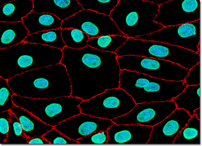
Immunohistochemistry is commonly distinguished as being the process of conducting immunoreactions on tissue thin sections, whereas immunocytochemistry is the same type of reactions on cell cultures. Furthermore, the term immunofluorescence is also often applied to immunochemical reactions that involve fluorophores conjugated to primary and secondary antibodies, as well as antibody fragments. In the scientific literature and the catalogs of antibody manufacturers, these terms are useful to segregate between products designed for cytological use and those targeted at paraffin or frozen tissue sections. However, the general principles of immunology are the same regardless of whether they are applied to isolated cells or those embedded within a tissue matrix. Simultaneous localization of tight junctions and the nuclear pore complex proteins (NPCP) was performed with a double immunofluorescence experiment with the LLC-PK1 cell culture illustrated above using mouse anti-NPCP and rabbit anti-ZO-3 primary antibodies. The subcellular targets were visualized using goat anti-mouse and anti-rabbit secondary antibodies (IgG) conjugated to Alexa Fluor 488 and Alexa Fluor 568, respectively. DNA in the nuclei was counterstained using Hoechst 33258. Images were recorded in grayscale with a QImaging Retiga Fast-EXi camera system coupled to an Olympus BX-51 microscope equipped with bandpass emission fluorescence filter optical blocks provided by Omega Optical. During the processing stage, individual image channels were pseudocolored with RGB values corresponding to each of the fluorophore emission spectral profiles. |
© 1995-2025 by Michael W. Davidson and The Florida State University. All Rights Reserved. No images, graphics, software, scripts, or applets may be reproduced or used in any manner without permission from the copyright holders. Use of this website means you agree to all of the Legal Terms and Conditions set forth by the owners.
This website is maintained by our
|