Fluorescence Digital Image Gallery
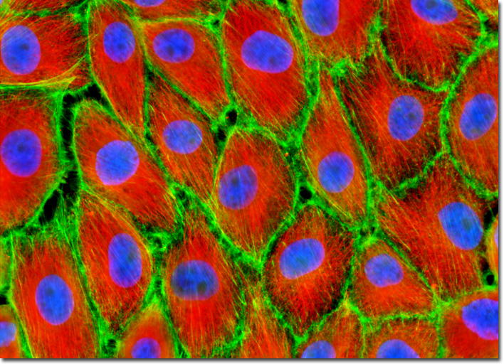
Pig Kidney Epithelial Cells (LLC-PK1)
|
The idea for immunofluorescence originated with Albert H. Coons and his collaborators, who were the first to conjugate a fluorescent dye to an antibody and use it identify a tissue antigen with fluorescence microscopy. The dye used in the original conjugation reaction was fluorescein isocyanate, but this species has now largely been replaced with the isothiocyanate derivative (fluorescein isothiocyanate, FITC), which forms a more stable conjugate. In most cases, when fluorescein is conjugated to an antibody or antibody fragment, the molecule fluoresces with a bright apple-green color after absorbing blue light in the region of 490 nanometers. A wide spectrum of alternative chromophores with fluorescent colors covering the entire visible and near-infrared spectrum are now commercially available as antibody conjugates, including the Alexa Fluor dyes, cyanine dyes (Cy2, Cy3, Cy5, etc.), and many rhodamine derivatives. An adherent culture of closely packed pig kidney epithelial cells, illustrated in the digital image presented above, was immunofluorescently stained with mouse anti-alpha-tubulin primary antibodies followed by goat anti-mouse Fab fragments conjugated to the cyanine dye, Cy3. The filamentous actin network was revealed by including Alexa Fluor 488 conjugated to phalloidin with the secondary antibody solution, and the nuclei were subsequently counterstained with DAPI. Images were recorded in grayscale with a QImaging Retiga Fast-EXi camera system coupled to an Olympus BX-51 microscope equipped with bandpass emission fluorescence filter optical blocks provided by Omega Optical. During the processing stage, individual image channels were pseudocolored with RGB values corresponding to each of the fluorophore emission spectral profiles. |
© 1995-2025 by Michael W. Davidson and The Florida State University. All Rights Reserved. No images, graphics, software, scripts, or applets may be reproduced or used in any manner without permission from the copyright holders. Use of this website means you agree to all of the Legal Terms and Conditions set forth by the owners.
This website is maintained by our
|