Fluorescence Digital Image Gallery
Pig Kidney Epithelial Cells (LLC-PK1)
|
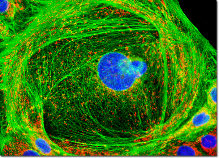
The proper functioning of the kidneys is extremely important to the body, which can suffer greatly if toxic substances are allowed to build-up in the blood. Humans can often lead a normal life if only one kidney is in good health, but if both of the organs are damaged, dialysis becomes necessary on a regular basis in order to filter the blood mechanically. Thus, medical understanding of the many renal diseases and disorders that have been identified is of considerable concern. The most common kidney problem reported is nephritis, a condition that usually occurs soon after other infections and which is characterized by the inflammation of glomeruli. Urinary tract infections and kidney stones are also frequently occurring renal conditions, but perhaps more important is the very serious, though less common, condition known as nephrosis. This degeneration of kidney tissue may result from a number of other disorders, including high blood pressure and diabetes, and is often not noticed until the condition is so advanced that kidney failure takes place and waste accumulation in the body becomes a dire circumstance. The adherent culture of LLC-PK1 epithelial cells featured in the digital image above was immunofluorescently labeled with primary anti-cytokeratin (pan) mouse monoclonal antibodies followed by goat anti-mouse Fab fragments conjugated to Alexa Fluor 488. In addition, the specimen was stained with MitoTracker Red CMXRos and DAPI, which bind with mitochondria and DNA, respectively. Images were recorded in grayscale with a QImaging Retiga Fast-EXi camera system coupled to an Olympus BX-51 microscope equipped with bandpass emission fluorescence filter optical blocks provided by Omega Optical. During the processing stage, individual image channels were pseudocolored with RGB values corresponding to each of the fluorophore emission spectral profiles. |
© 1995-2025 by Michael W. Davidson and The Florida State University. All Rights Reserved. No images, graphics, software, scripts, or applets may be reproduced or used in any manner without permission from the copyright holders. Use of this website means you agree to all of the Legal Terms and Conditions set forth by the owners.
This website is maintained by our
|