Fluorescence Digital Image Gallery
Human Cervical Adenocarcinoma Cells (HeLa)
|
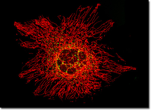
Several plasmids designed to localize enhanced green fluorescent protein (EGFP) to the peroxisomes have been constructed and employed to reliably transfect susceptible mammalian cell lines. One commercially available plasmid, designated EGFP-Peroxi, encodes a fusion protein consisting of the EGFP nucleotide sequence and the peroxisomal targeting signal 1 (PTS1). The PTS-1 sequence, which transports the chimeric protein (EGFP and peroxisomal signal) to the matrix of the peroxisomes, is fused to the 3' terminus of the EGFP sequence and encodes the tripeptide serine-lysine-leucine. This version of enhanced green fluorescent protein nucleotide sequence produces a red-shifted mutant of the wild-type that has been optimized for brighter fluorescence, with an absorption maximum at 488 nanometers (designed to coincide with a principal argon-ion laser spectral line) and an emission maximum wavelength of approximately 507 nanometers. Illustrated in the digital image above is a resident from a culture of HeLa cervical carcinoma epithelial cells that were transfected with an EGFP vector containing the peroxisomal targeting signal 1 (PTS1) fusion protein. Stable transfectants were isolated and subsequently labeled with MitoTracker Red CMXRos before being counterstained with Hoechst 33342. The specimen was imaged using a FluoView FV300 scanning confocal microscope equipped with an argon-ion and a helium-neon laser (488 and 543 nanometer excitation wavelengths, respectively). |
© 1995-2025 by Michael W. Davidson and The Florida State University. All Rights Reserved. No images, graphics, software, scripts, or applets may be reproduced or used in any manner without permission from the copyright holders. Use of this website means you agree to all of the Legal Terms and Conditions set forth by the owners.
This website is maintained by our
|