Fluorescence Digital Image Gallery
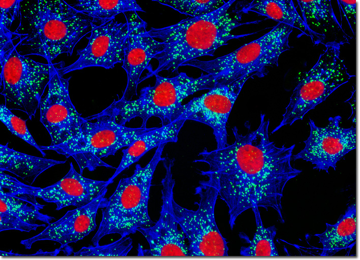
Transformed Chicken Embryo Fibroblast Cells (UMNSAH/DF-1)
|
Similar to mitochondria, peroxisomes are heavily involved in cellular metabolic processes. The ubiquitous organelles, which are delineated by a single membrane, generally contain enzymes that utilize oxygen to subtract hydrogen atoms from certain organic substrates in an oxidative reaction that generates hydrogen peroxide. Peroxisomes also typically contain catalase, an enzyme that uses this toxic byproduct of metabolism to oxidize formic acid, alcohols, phenols, and other substrates, a function that is especially important in cells of the kidneys and liver, which are responsible for the detoxification of various toxins traveling through the bloodstream. Any remaining hydrogen peroxide present in the cell is broken down by catalase into water and free oxygen molecules. The degradation of fatty acids and the catalysis of the initial steps in the synthesis of ether phospholipids, which are eventually utilized in membrane formation, are a few of the other various tasks commonly carried out by peroxisomes. In a double immunofluorescence experiment, the log phase adherent monolayer culture of chicken embryo fibroblast cells illustrated above was fixed, permeabilized, blocked with 10 percent normal goat serum, and treated with a cocktail of mouse anti-histones (pan) and rabbit anti-PMP 70 (peroxisomal membrane protein) primary antibodies, followed by goat anti-mouse and anti-rabbit secondary antibodies (IgG) conjugated to Texas Red and Alexa Fluor 488, respectively. The filamentous actin network was counterstained with Alexa Fluor 350 conjugated to phalloidin. Images were recorded in grayscale with a QImaging Retiga Fast-EXi camera system coupled to an Olympus BX-51 microscope equipped with bandpass emission fluorescence filter optical blocks provided by Omega Optical. During the processing stage, individual image channels were pseudocolored with RGB values corresponding to each of the fluorophore emission spectral profiles. |
© 1995-2025 by Michael W. Davidson and The Florida State University. All Rights Reserved. No images, graphics, software, scripts, or applets may be reproduced or used in any manner without permission from the copyright holders. Use of this website means you agree to all of the Legal Terms and Conditions set forth by the owners.
This website is maintained by our
|
