Fluorescence Digital Image Gallery
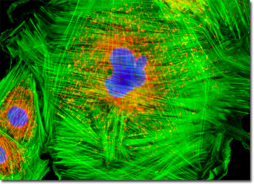
Embryonic Rat Thoracic Aorta Medial Layer Myoblast Cells (A-10)
|
Found primarily lining the hollow organs of the body, such as the thoracic aorta from which A-10 cells were derived, smooth muscle cells are usually arranged in dense sheets. The cells of the sheets are interconnected by gap junctions, specialized communication ports located between the cells. These junctions are composed of arrays of small channels that permit small molecules to shuttle from one cell to another, essentially linking the internal environment of one cell with those of adjacent cells. The force generating ability of smooth muscle is greater than that of other muscle types and the amount of time the force may be maintained is also significantly greater. The regulation of smooth muscle activity involves either the autonomic nervous system or hormones in the blood system. In some cases, smooth muscle tissue reacts to both types of regulation. The culture of A-10 cells illustrated above was labeled with MitoTracker Red CMXRos before fixing, and the cells were subsequently stained with Alexa Fluor 488 conjugated to phalloidin, followed by Hoechst 33258, a popular DNA-binding counterstain. Images were recorded in grayscale with a QImaging Retiga Fast-EXi camera system coupled to an Olympus BX-51 microscope equipped with bandpass emission fluorescence filter optical blocks provided by Omega Optical. During the processing stage, individual image channels were pseudocolored with RGB values corresponding to each of the fluorophore emission spectral profiles. |
© 1995-2025 by Michael W. Davidson and The Florida State University. All Rights Reserved. No images, graphics, software, scripts, or applets may be reproduced or used in any manner without permission from the copyright holders. Use of this website means you agree to all of the Legal Terms and Conditions set forth by the owners.
This website is maintained by our
|