Fluorescence Digital Image Gallery
Embryonic Rat Thoracic Aorta Medial Layer Myoblast Cells (A-10)
|
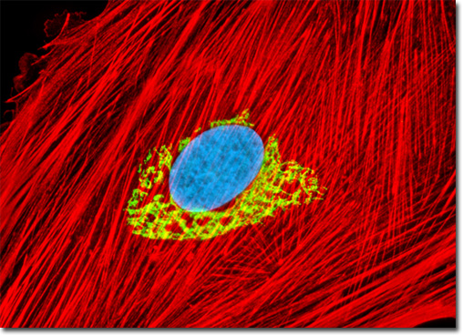
The Golgi apparatus present in mammalian cells may be labeled with immunofluorescent probes, but first a candidate protein resident somewhere in the complex must be identified and targeted with a primary mono or polyclonal antibody. Giantin is a membrane-bound glycoprotein that resides in the cis and medial regions of the Golgi apparatus, which contains an elongated rod-like cytoplasmic domain 350 kiloDaltons or more in size. Research suggests that the highly conserved protein may be involved in intercisternal cross-bridge structures that are assumed to function in Golgi cisternae stacking. Polyclonal antibodies raised against the N-terminal 469 peptide residues of giantin react with a number of mammalian species. Also, monoclonal antibodies have been produced that detect the protein in avian specimens. The culture of rat thoracic aorta cells illustrated above was fixed, permeabilized, and blocked with 10-percent normal goat serum in phosphate-buffered saline prior to immunofluorescent labeling with rabbit primary antibodies to giantin, a protein resident in the Golgi complex of mammalian cells. The culture was subsequently stained with a mixture of goat anti-rabbit secondary antibody fragments (heavy and light chain) conjugated to Cy2. In addition, the cells were labeled with Alexa Fluor 568 conjugated to phalloidin (targeting the filamentous actin network), and for DNA with Hoechst 33342. Images were recorded in grayscale with a QImaging Retiga Fast-EXi camera system coupled to an Olympus BX-51 microscope equipped with bandpass emission fluorescence filter optical blocks provided by Omega Optical. During the processing stage, individual image channels were pseudocolored with RGB values corresponding to each of the fluorophore emission spectral profiles. |
© 1995-2025 by Michael W. Davidson and The Florida State University. All Rights Reserved. No images, graphics, software, scripts, or applets may be reproduced or used in any manner without permission from the copyright holders. Use of this website means you agree to all of the Legal Terms and Conditions set forth by the owners.
This website is maintained by our
|