Fluorescence Digital Image Gallery
Embryonic Rat Thoracic Aorta Medial Layer Myoblast Cells (A-10)
|
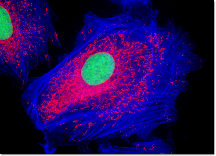
Due to the significant amount of elastic material it exhibits, the aorta is known as an elastic artery. Under the microscope, special stains are often required, however, in order to readily observe the elastic material. The main branches of the aorta as well as the other largest arteries of the body, which include the pulmonary artery and its major branches, are also considered elastic. All of these important blood vessels have a similar functional role, ensuring that blood is transported smoothly and continuously through the tubes. Their ability to carry out this function depends heavily upon the pumping of the heart, which pushes blood into the elastic arteries while contracting. The pressure that results transports blood along the elastic arteries and simultaneously causes them to distend, an action chiefly enabled by the significant amount of elastic material they contain. When the heart relaxes, however, pressure is removed from the arteries and their subsequent shrinkage serves to maintain blood pressure within the arteries and a constant flow of blood through the circulatory system. The culture of adherent A-10 rat thoracic aorta cells presented in the digital image above was fluorescently triple-labeled with MitoTracker Red CMXRos, Alexa Fluor 350 conjugated to phalloidin, and SYTOX Green, targeting the mitochondria, filamentous actin network, and nuclei, respectively. In this image, the bright red mitochondrial network is superimposed on a deep blue actin cytoskeletal framework centered around the green nuclei. Images were recorded in grayscale with a QImaging Retiga Fast-EXi camera system coupled to an Olympus BX-51 microscope equipped with bandpass emission fluorescence filter optical blocks provided by Omega Optical. During the processing stage, individual image channels were pseudocolored with RGB values corresponding to each of the fluorophore emission spectral profiles. |
© 1995-2025 by Michael W. Davidson and The Florida State University. All Rights Reserved. No images, graphics, software, scripts, or applets may be reproduced or used in any manner without permission from the copyright holders. Use of this website means you agree to all of the Legal Terms and Conditions set forth by the owners.
This website is maintained by our
|