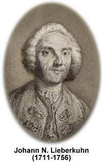
Johann Nathanael Lieberkühn
(1711-1756)

Johann Nathanael Lieberkühn was a German physician, anatomist, and physicist. He is most widely known for development of the solar microscope, studies of the intestine, and invention of a reflector for improving microscopic viewing of opaque specimens. He was also a member of the mathematics department at the Berlin Academy of Sciences and created a lens that enhanced the use of early portable microscopes for botanical fieldwork.
During his lifetime, normal and pathological anatomies were more established sciences than microscopy. Yet, Lieberkühn's work heavily depended on the microscope. As an anatomist, his research largely focused on the digestive system. The crypts of Lieberkuhn, named in his honor, are the intestinal glands found between the villi that act as a source for digestive enzymes and various hormones. The receptors and carriers necessary for digestion and absorption are contained in the complex network of membranes. Some of Lieberkühn's anatomical specimens have survived for over 250 years. When Catherine the Great acquired them in 1765, she placed them in the Russian Medical Military Academy's Museum of Anatomy, where they remain today.
To aid his anatomical work, Lieberkühn used a simple microscope fitted with a concave mirror or reflector to study injected animal specimens with epi-illumination. The Lieberkuhn reflector, or reflecting speculum, is made of silver or another highly polished metal, and increases the amount of light illuminating a specimen. The main advantage of the reflector is that it illuminates an opaque object from almost every azimuth. Although used by many early microscope makers, for most applications the stereomicroscope and modern incident lighting have made the Lieberkuhn device obsolete. Olympus, however, still manufacturers Lieberkühn's design as an option for some of its modern microscope models that use 20-millimeter and 38-millimeter macro lenses.
Lieberkühn invented the solar microscope around 1740. Structurally similar to a microscope, in reality it was a projector that required very intense light for good resolution of fine anatomical details. The microscope was designed for showing magnifications of transparent specimens to large audiences, such as an anatomy class. The horizontal instrument was placed at the base of a window along with a mirror that collected and reflected the incident sunlight. A bi-convex lens focused the light rays onto the transparent specimen. The resulting image was enlarged and could be projected a considerable distance from the instrument and object. If sunlight was not accessible, a gas lamp or other light source could alternatively be used with the microscope.
