
Oblique Transmitted Illumination
Oblique illumination, both through the microscope and in everyday life, enables light to strike an object at a low glancing angle with a resulting shadow effect on one side of the object. The other side is illuminated with the oblique light rays to produce an apparent relief detail on the object that stands out with three-dimensional clarity.
In the microscope, the technique of oblique illumination produces a direct light that is restricted to a single azimuth of the condenser light cone, and is allowed to illuminate the specimen from one side only. The net effect is to reveal details in an otherwise almost invisible, colorless specimen in pseudo-relief.
The oblique lighting forces the zeroth order (undeviated) light rays to be moved to a position just within the periphery of the objective. In research-grade microscopes this can be observed with a focusing telescope (or Bertrand lens) at the rear focal plane of the objective, an area not readily imaged with the QX3 computer microscope. Shifting of the zeroth order light rays to the periphery allows additional diffracted orders (or sometimes just a single diffracted order) to be included in the objective's rear focal plane. Higher order diffracted oblique light rays appear on one side of the objective only, because diffracted orders on the other side fail to enter the objective front lens.
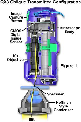
Adaptation of the QX3 microscope for oblique illumination requires a substage condenser with a slit aperture strategically placed at the periphery of the condenser base, as illustrated in Figure 1. In this configuration, a Hoffman-style condenser containing slits of varying sizes is used to provide oblique illumination with controllable intensity. Slit size in this type of condenser is matched to individual objective requirements, with the largest slits designed for the lower power objectives, and visa versa. Use of a condenser to achieve oblique illumination is absolutely essential, because light intensity from the stock QX3 mixing chamber and diffusion screen is insufficient to illuminate specimens from an oblique angle, even with the assistance of auxiliary illumination.
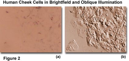
The result of oblique transmitted illumination is to increase apparent resolution and also to produce a shadowed, relief-like pseudo three-dimensional appearance in the image of the specimen. This method works well with many stained and unstained objects such as living cells, crystals, diatoms, rock thin sections, etc. The resulting image must be viewed with caution because the diffracted orders from one side have not contributed to formation of the image.
A simpler method of obtaining oblique transmitted illumination is to decenter the substage condenser aperture diaphragm. In former years, several microscopes were equipped with a condenser having a decenterable aperture iris diaphragm. The device was engineered to allow the entire iris to move off-center in a horizontal plane such that closing the iris disphragm would result in moving the zeroth order light rays to the periphery of the objective rear focal plane. This method is similar to that described above for the Hoffman-style condenser. The entire diaphragm was rotatable around the axis of the microscope so that oblique light could be directed toward the specimen from any azimuth to achieve the best desired effect for a given specimen. Oblique transmitted illumination is a very good example of how manipulation of light at the level of the aperture iris diaphragm (at the front focal plane of the condenser) can significantly alter the appearance of the image at the position of the eyepiece fixed diaphragm or the QX3 CMOS photodiode array.
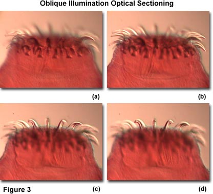
Another advantage of using oblique illumination is the ability to perform "optical sectioning" of transparent specimen with this technique. Sectioning allows the microscopist to focus on a single thin plane of the specimen without interference from confusing images arising in areas above or below the plane that is being focused on. The depth of a specimen is measured in a direction parallel to the optical axis of the microscope. Focusing the image establishes the correct specimen-to-image distance, allowing interference of the diffracted waves to occur at a pre-determined plane (the image plane) positioned at a fixed distance from the eyepiece. This enables diffracting objects that occur at different depth levels in the specimen to be viewed separately, provided there is sufficient contrast. The entire depth of a specimen can be optically sectioned by sequentially focusing on each succeeding plane. In this system, depth of field is defined as the distance from one level to the next where imaging of distinct detail occurs, and is controlled by the numerical aperture of the objective. Higher numerical aperture objectives exhibit very shallow depths of field and the opposite holds for objectives of lower numerical aperture. The overall capability of an objective to isolate and focus on a specific optical section diminishes as the optical homogeneity of the specimen decreases.
BACK TO INTEL QX3 SPECIALIZED TECHNIQUES
Questions or comments? Send us an email.
© 1995-2025 by Michael W. Davidson and The Florida State University. All Rights Reserved. No images, graphics, software, scripts, or applets may be reproduced or used in any manner without permission from the copyright holders. Use of this website means you agree to all of the Legal Terms and Conditions set forth by the owners.
This website is maintained by our
Graphics & Web Programming Team
in collaboration with Optical Microscopy at the
National High Magnetic Field Laboratory.
The QX3 microscope design is copyrighted © 2025 by Mattel, Inc. Intel® Play™ is a registered trademark of the Intel Corporation. These companies reserve all of their rights and privileges under copyright law. The material contained in this website is solely the opinion of the authors and is intended for eduational use only.
Last Modification Friday, Nov 13, 2015 at 02:18 PM
Access Count Since April 22, 2000: 33513
Visit the official Intel Play website:
![]()