
Advanced Condenser Systems: Achromatic Condensers
Uracil Crystallites
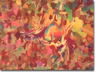
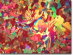
The images below compare performance of the Intel Play QX3 Computer Microscope with and without the aid of an organized cone of illumination from an achromatic substage condenser containing an aperture diaphragm. These photomicrographs are unretouched and were captured with the QX3 interactive software.
Birefringent Uracil Crystallites
Dihydroxy Pyrimidine, or uracil, is a crystalline organic compound found in ribonucleic acid (RNA) and is also a component of coenzymes that work with enzymes in carbohydrate metabolism. Like deoxyribonucleic acid (DNA), RNA is composed of a series of nucleotides, each containing one of four nitrogen-containing bases. However, while the bases adenine, guanine, and cytosine are found in both DNA and RNA, RNA uses uracil instead of the base thymine, which is found in DNA.
RNA functions as hereditary material in viruses and performs protein synthesis in cells since the DNA is unable to make protein. There are three types of cellular RNA: messenger RNA (mRNA), transfer RNA, and ribosomal RNA (rRNA). To manufacture proteins, mRNA carries codes from DNA in the nucleus to the ribosomes in the cytoplasm. In the cytoplasm, it builds amino acids, which the tRNA brings to the rRNA in the ribosomes to be linked together into proteins.
RNA also occurs in a number of other forms inside cells, as small RNA molecules and molecules composed of RNA and protein. These forms of RNA appear to act as biological catalysts, a function previously thought to belong solely to proteins.
The images above were recorded using the Intel Play QX3 microscope in transmitted brightfield mode equipped with crossed polarizers and a full-wave retardation plate. On the top is a digital image from a stock QX3 microscope using auxiliary illumination provided by a fiber optic light pipe through a hole drilled into the mixing chamber. The image on the bottom was recorded using the QX3 microscope body and a Nikon achromatic substage condenser of low numerical aperture.
BACK TO TRANSMITTED POLARIZED ILLUMINATION GALLERY
Questions or comments? Send us an email.
© 1995-2025 by Michael W. Davidson and The Florida State University. All Rights Reserved. No images, graphics, software, scripts, or applets may be reproduced or used in any manner without permission from the copyright holders. Use of this website means you agree to all of the Legal Terms and Conditions set forth by the owners.
This website is maintained by our
Graphics & Web Programming Team
in collaboration with Optical Microscopy at the
National High Magnetic Field Laboratory.
The QX3 microscope design is copyrighted © 2025 by Mattel, Inc. Intel® Play™ is a registered trademark of the Intel Corporation. These companies reserve all of their rights and privileges under copyright law. The material contained in this website is solely the opinion of the authors and is intended for eduational use only.
Last Modification Friday, Nov 13, 2015 at 02:19 PM
Access Count Since April 25, 2000: 11495
Visit the official Intel Play website:
![]()