
Advanced Condenser Systems: Abbe Condensers
Oblique Transmitted (Hoffman-Style) Illumination
Human Cheek Epithelial Cells
The skin that lines the mouth is called the buccal muscosa and is composed of squamous epithelial cells that divide about once every 24 hours. As evidenced by these micrographs, oblique illumination adds a pseudo three-dimensional effect to images when compared to unaided brightfield microscopy.
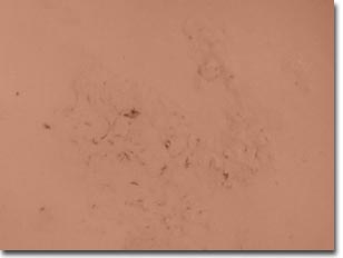
(Brightfield)
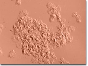
(Oblique Illumination)
These cells secrete mucin, a mucopolysaccharide that is the principal constituent of mucus, which helps keep the interior of the mouth moist in addition to the salivary glands. The individual cells have a flat, irregular shape and a very thin membrane. Cheek cells are a popular item of study in the classroom. These easily obtained specimens are viewed under the microscope and used as a source for DNA fingerprinting studies. They are also used in diagnostic tests. Children born with "ambiguous" genitalia can have their sex determined by a simple scraping of cheek cells and a test for the presence of Barr Bodies (inactivated X chromosomes generally found only in women). In Australia, a test using cheek cells has been devised that can measure a person's proclivity for high blood pressure.
Transmitted oblique illumination was obtained with a Hoffman-style substage condenser fitted with an offset aperture slit, which provides illumination from an oblique angle with respect to the optical axis of the QX3 microscope. Digital images were recorded with the QX3 interactive software operating from the Live View menu. In the case of birefringent specimens, a polarizer was added beneath the Hoffman condenser and an analyzer was placed in front of the QX3 lens cover. In some instances, a full-wave retardation plate was inserted between the specimen and the analyzer before capturing images.
BACK TO THE OBLIQUE (HOFFMAN) GALLERY
Questions or comments? Send us an email.
© 1995-2025 by Michael W. Davidson and The Florida State University. All Rights Reserved. No images, graphics, software, scripts, or applets may be reproduced or used in any manner without permission from the copyright holders. Use of this website means you agree to all of the Legal Terms and Conditions set forth by the owners.
This website is maintained by our
Graphics & Web Programming Team
in collaboration with Optical Microscopy at the
National High Magnetic Field Laboratory.
The QX3 microscope design is copyrighted © 2025 by Mattel, Inc. Intel® Play™ is a registered trademark of the Intel Corporation. These companies reserve all of their rights and privileges under copyright law. The material contained in this website is solely the opinion of the authors and is intended for eduational use only.
Last Modification Friday, Nov 13, 2015 at 02:19 PM
Access Count Since April 26, 2000: 68274
Visit the official Intel Play website:
![]()