Advanced Condenser Systems: Achromatic Condensers
Mosquito Whole Mount
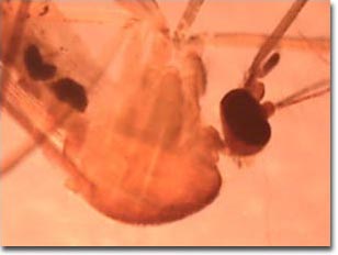
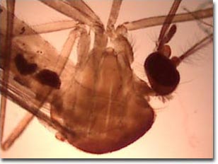
Semi-transparent insects are difficult to image using unaided brightfield optical microscopy. The images below compare performance of the Intel Play QX3 Computer Microscope with and without the aid of an organized cone of illumination from a substage condenser containing an aperture diaphragm. These photomicrographs are unretouched and were captured with the QX3 interactive software.
Unstained Mosquito Whole Mount


The Culex genus of mosquitoes has a worldwide distribution and is found on every continent except Antarctica. Fourteen species occur throughout the United States and Canada. Many species of Culex are known as the "house mosquito" because these insects commonly develop in small containers around houses. The main host for Culex is wild birds, but it also feeds on a wide variety of warm-blooded vertebrates, including humans.
Culex is a carrier of viral encephalitis and, in tropical and subtropical climates, of filariasis. In the US, they are disease vectors for western equine and Saint Louis encephalitis. They are also vectors for the West Nile virus that suddenly appeared in the Queens borough of New York City in the summer of 1999, killing six people. The virus, established in Africa and Europe, was most likely introduced into the US by accident. When exactly this happened is subject to debate. The virus may have been in the US for years, but until people started dying from it, one knew about it. Given that Culex and birds are found everywhere there are people in North America, this disease is probably here to stay.
The images above were recorded using the Intel Play QX3 microscope in transmitted brightfield mode. On the top is a digital image from a stock QX3 microscope using auxiliary illumination provided by a fiber optic light pipe through a hole drilled into the mixing chamber. The image on the bottom was recorded using the QX3 microscope body and a Nikon achromatic substage condenser with low numerical aperture. Note the ability of the microscope to resolve tiny hairs on the insect's body, legs, head, and antennae when illumination is organized and controlled by a condenser.
BACK TO TRANSMITTED BRIGHTFIELD GALLERY
