Advanced Condenser Systems: Abbe Condensers
Cartilage
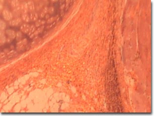
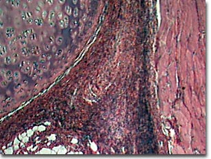
The images below compare performance of the Intel Play QX3 Computer Microscope with and without the aid of an organized cone of illumination from a substage condenser containing an aperture diaphragm. The digital images are unretouched and were captured with the QX3 interactive software.
Cartilage is a dense network of collagen fibers, embedded in a firm but flexible plastic-like substance. It forms the skeletons of young mammals, before bone starts forming, and persists in parts of the skeleton into adulthood. Primitive vertebrates, such as sharks and lampreys, have skeletons composed only of cartilage.
Cartilage Whole Mount


(200x magnification)
Semi-transparent and lightly stained specimens are often very difficult to image using unaided brightfield optical microscopy. The images presented here were recorded using the Intel Play QX3 microscope in transmitted brightfield mode. On the top is a digital image from a stock QX3 microscope using either auxiliary illumination provided by a fiber optic light pipe through a hole drilled into the mixing chamber, or standard illumination from the microscope's tungsten lamp and mixing chamber. The image on the bottom was recorded using the QX3 microscope body coupled to a simple two-lens Abbe low numerical aperture substage condenser. Illumination was provided by a 30 watt tungsten bulb housed in an illuminator with a heat sink, a frosted diffusion screen, and a daylight color-compensating filter.
BACK TO ABBE CONDENSER BRIGHTFIELD GALLERY
