Observing Mitosis with Fluorescence Microscopy
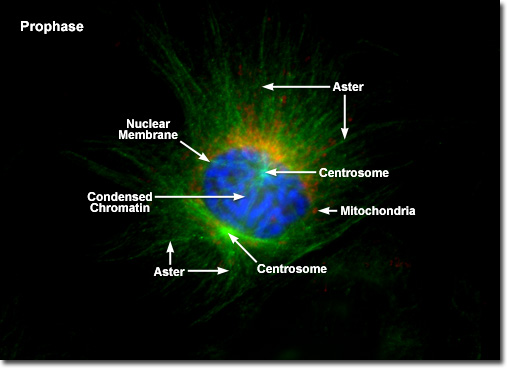
Prophase
|
In order to ensure the inheritance of a complete ensemble of critical internal components by each daughter cell, the cell division process must provide for the segregation of organelles, such as mitochondria, the Golgi apparatus, and endoplasmic reticulum during mitosis. For example, the tubular, self-contained mitochondria are unable to assemble spontaneously from their component proteins, lipids, and enzymes, and can only be duplicated by growth and fission of pre-existing organelles. In a similar manner, the large interconnected membranous complexes of the endoplasmic reticulum and Golgi apparatus require at least a portion of the original structure as a master template for duplication of the entire network. Segregation is accomplished by doubling the concentration of self-contained organelles (mitochondria) or by fragmentation, followed by vesicle formation, of the larger membrane-rich organelles (endoplasmic reticulum) during cell division. Vesicles of the endoplasmic reticulum and Golgi apparatus appear to associate to some degree with the mitotic spindle, which is probably a mechanism designed to ensure even distribution between the daughter cells. |
© 1995-2025 by Michael W. Davidson and The Florida State University. All Rights Reserved. No images, graphics, software, scripts, or applets may be reproduced or used in any manner without permission from the copyright holders. Use of this website means you agree to all of the Legal Terms and Conditions set forth by the owners.
This website is maintained by our
|