|
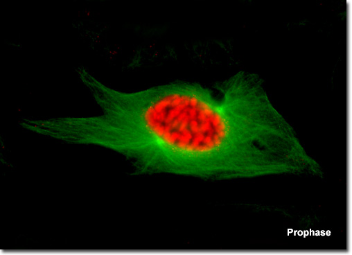
Presented in the digital fluorescence microscopy image above is a single kangaroo rat (PtK2) kidney cell in the early stages of prophase. The chromatin is stained with a red fluorescent probe (Alexa Fluor 568), while the microtubule network (mitotic spindle) is stained green (Alexa Fluor 488). The mitotic spindle that begins to form in early prophase is a bipolar structure composed of microtubules and associated proteins. As the spindle grows, the centrosomes begin to translocate to opposite ends of the nucleus, apparently driven by the addition of new tubulin monomers to the existing filament network. On the molecular level, three sets of spindle microtubules can be distinguished. The polar microtubules overlap in the central region of the spindle and are responsible for pushing the poles (centrosomes) farther apart. Kinetochore microtubules attach to a specialized region within the centromeres of the sister chromatids to assist in maneuvering the chromosomes to the mitotic plate. Radiating in all directions from the centrosomes at the spindle poles are astral microtubules, which separate the poles and orient the mitotic spindle with respect to other cellular components.
|
