Observing Mitosis with Fluorescence Microscopy
Prometaphase
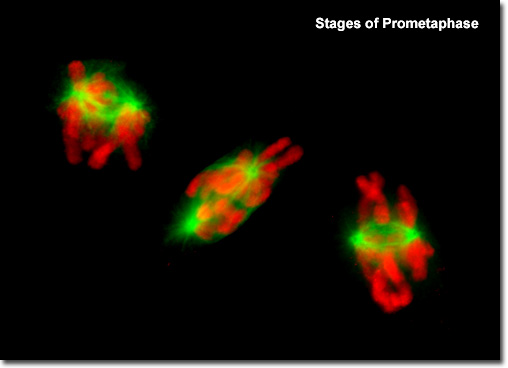
|
Presented in the digital fluorescence microscopy image above are rat kangaroo (PtK2) kidney epithelial cells in various stages of prometaphase. The chromatin is stained with a bright red fluorescent probe (Alexa Fluor 568), while the microtubule network (mitotic spindle) is stained green (Alexa Fluor 488). During prometaphase, the mitotic spindle microtubules are now free to enter the nuclear region, and formation of specialized protein complexes known as kinetochores begins on each centromere. These complexes become attached to a subset of the spindle microtubules, which are then termed kinetochore microtubules. Other microtubules in the spindle (not attached to centromeres) are termed polar microtubules, and these help form and maintain the spindle structure along with astral microtubules, which remain outside the spindle. |
© 1995-2025 by Michael W. Davidson and The Florida State University. All Rights Reserved. No images, graphics, software, scripts, or applets may be reproduced or used in any manner without permission from the copyright holders. Use of this website means you agree to all of the Legal Terms and Conditions set forth by the owners.
This website is maintained by our
|