Observing Mitosis with Fluorescence Microscopy
Interphase
|
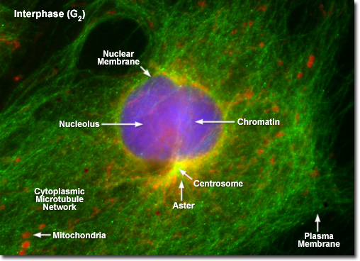
Illustrated in the fluorescence digital image above is a rat kangaroo (PtK2) kidney cell in the second gap (G(2)) period of interphase. The chromatin is stained with a blue fluorescent probe (DAPI), while the microtubule network is stained green (Alexa Fluor 488) and cellular mitochondria are stained with a red dye (MitoTracker Red CMXRos). The centrosomes act as the principal microtubule organizing center for the cell to nucleate a cytoplasmic array of microtubules, which project outward towards the plasma membrane. During interphase, the centrioles and other components of the centrosomal matrix are duplicated, but remain complexed into a single unit on one side of the nucleus, as presented in the image. At the onset of mitosis, the centrosome complex divides with each centriole pair becoming the center of an organized network of radial microtubules. |
© 1995-2025 by Michael W. Davidson and The Florida State University. All Rights Reserved. No images, graphics, software, scripts, or applets may be reproduced or used in any manner without permission from the copyright holders. Use of this website means you agree to all of the Legal Terms and Conditions set forth by the owners.
This website is maintained by our
|