Observing Mitosis with Fluorescence Microscopy
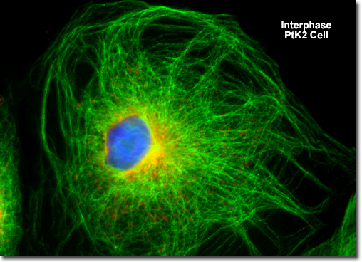
Interphase
|
Although very little activity is observable in the cell nucleus with fluorescence microscopy during interphase and the period is not considered to be a formal step in mitosis, this stage represents an essential preparation for cell division because the chromosomes are replicated during interphase. In addition to the synthesis phase, which occurs during the central portion of interphase, the cell cycle also consists of two gap (abbreviated G) stages that precede and follow the synthesis phase. For most animal cells, the interphase portion accounts for approximately 90 percent of the cell cycle, whereas mitosis is accomplished in the remaining period. |
© 1995-2025 by Michael W. Davidson and The Florida State University. All Rights Reserved. No images, graphics, software, scripts, or applets may be reproduced or used in any manner without permission from the copyright holders. Use of this website means you agree to all of the Legal Terms and Conditions set forth by the owners.
This website is maintained by our
|