Observing Mitosis with Fluorescence Microscopy
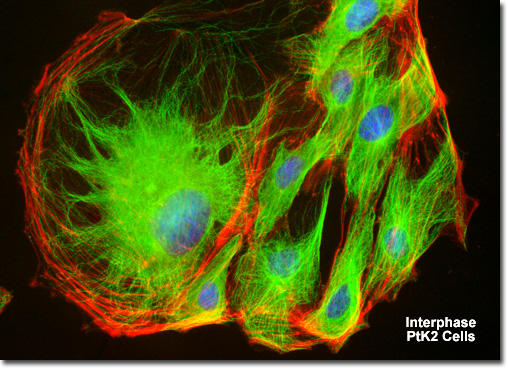
Interphase
|
When observed through the microscope, the cell nucleus is well defined and surrounded by the nuclear envelope (or membrane) during interphase. Within the nuclear membrane are one or more nucleoli, which appear as spherical, dense structures when stained with fluorescent or absorbing dyes. On the outer periphery of the nucleus are two important organelles termed centrosomes, which were formed by duplication of a single copy during the last division cycle. Each centrosome contains a pair of centrioles that extrude visible microtubule networks through the cytoplasm in radial arrays, known as asters (for stars). Even though the chromosomes have been duplicated during the DNA synthesis (S) phase, individual chromatids are not visible in late interphase because the chromosomes still exist in the form of loosely packed chromatin fibers. |
© 1995-2025 by Michael W. Davidson and The Florida State University. All Rights Reserved. No images, graphics, software, scripts, or applets may be reproduced or used in any manner without permission from the copyright holders. Use of this website means you agree to all of the Legal Terms and Conditions set forth by the owners.
This website is maintained by our
|