
Polarized Light Microscopy Digital Image Gallery
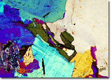
Biotite in Granite
Granite is the most widespread igneous rock comprising the crust of the Earth. Commonly quarried, the visibly crystalline, light-colored rock has been utilized as a paving and building material since antiquity.
View a second image of Biotite in Granite
A variety of classes of granites have been established by geologists, most of which are differentiated by the relative coarseness of the grain or the amounts of the constituent minerals that are present. Granites are, however, chiefly composed of plagioclase and alkali feldspars. Other minor minerals that are commonly incorporated in the rocks include muscovite, pyroxenes, and biotite. Indeed, biotite is almost always found in granites of all types, though sometimes in negligible quantities.
Biotite is one of the most abundant members of the group of minerals known as micas. In fact, the mineral, which varies somewhat in color though it is usually quite dark, is sometimes alternatively referred to as black mica. While frequently found in igneous and metamorphic rocks, biotite’s tendency to easily alter when subjected to chemical weathering makes the mineral a rare occurrence in sedimentary rocks. Interestingly, in a stage of partial alteration, biotite lightens in color and has an appearance similar to metallic flakes, which are occasionally misidentified by the casual observer as flecks of gold.