
Phase Contrast Image Gallery
Bronchial Pneumonia
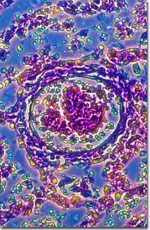
A stained thin section of human lung tissue exhibiting damage from bronchial pneumonia is illustrated below. As evidenced by the micrograph, combining phase contrast microscopy with classical histological staining techniques in pathological research often yields enhancement of cellular features.
Bronchopneumonia, or bronchial pneumonia, is a contagious infection of the lungs caused by bacteria, viruses, or other microorganisms. This type of pneumonia affects the smaller branches of the bronchial tubes, called bronchioles, which become inflamed as they clog with pus and mucus.
Before antibiotic drugs were developed in the 1940's, pneumonia killed about one-third of its victims. Now over 95 percent of all patients recover with proper medical treatment. Nonetheless, pneumonia still ranks as one of the top ten leading causes of death in the United States.
Influenza viruses and streptococcal bacteria are common causes of pneumonia, although they usually have the greatest affect on those who are already ill. Patients with seriously compromised immune systems can develop pneumonia from organisms that are not normally pathogenic (disease causing).
Some virulent forms of pneumonia-causing pathogens, such as Legionnaire's Disease, can affect otherwise healthy people. In 1976, an outbreak of severe pneumonia occurred among U.S. veterans attending a convention of the American Legion in Philadelphia. The bacterium, later named Legionella pneumophilia, infected 147 people and killed 29. It had not been detected previously because it didn't show up with the type of laboratory tests that were being used to find most pneumococci (bacteria that cause acute pneumonia). L. pneumophilia can grow in air-conditioning systems or on showerheads, and has been shown to be responsible for sporadic but severe outbreaks of pneumonia. Now that it can be detected, however, it is easily treated with conventional antibiotics.
BACK TO THE PHASE CONTRAST GALLERY