
Fluorescence Digital Image Gallery
Obelia Hydroid: First Generation
Obelia belongs to the phylum Cnidaria, which includes corals, sea anemones, jellyfish, and the freshwater hydra. The many species of this genus are widely distributed throughout all the oceans and are typical of cnidarians, both in their morphology and their life cycle.
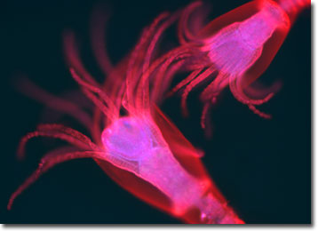
These animals take two generations to complete one life cycle. One generation, illustrated in the photomicrograph above, lives in hydroid colonies, consisting of polyps. The polyps are stalklike forms that attach to a surface (usually the ocean bottom) by means of root-like filaments. The polyps reproduce asexually, by budding, and create new polyps until it has formed a treelike colony. The colonies are dimorphic, having two types of polyps. Gastrozooids, or hydranths, are the feeding polyps. They have a mouth surrounded by stinging tentacles, giving them a flower-like appearance, and are responsible for capturing and consuming food. Food is digested in the gastrovascular cavity and provided to the rest of the colony. Gonozooids are the reproductive polyps and, through budding, produce the next generation--tiny jellyfish called medusae.
The second generation of the Obelia life cycle begins when the medusae are released by the gonozooids and become free-swimming forms. The medusae reproduce sexually, producing eggs and sperm that fertilize to become ciliated larvae, known as planulae. The planulae remain in a free-swimming form for a period of time, eventually attaching to a surface and developing into polyps.
The specimen presented here was imaged with a Nikon E600 microscope operating with fluorite and/or apochromatic objectives and vertical illuminator equipped with a mercury arc lamp. Specimens were illuminated through Nikon dichromatic filter blocks containing interference filters and a dichroic mirror and imaged with standard epi-fluorescence techniques. The filter combination utilized for the obelia stained specimen was a triple cube specific for DAPI, FITC, and Texas Red. Photomicrographs were captured with a Optronics MagnaFire digital camera system coupled to the microscope with a lens-free C-mount adapter.
BACK TO THE FLUORESCENCE DIGITAL IMAGE GALLERY