
Fluorescence Digital Image Gallery
Normal African Green Monkey Kidney Fibroblast Cells (CV-1 Line)
The CV-1 cell line was initiated in March of 1964 by F. C. Jensen and his colleagues with a tissue section excised from the kidney of a normal adult male African green monkey (Cercopithecus aethiops). The popular fibroblast line was originally utilized in research focusing on the transformation of the cancer-causing Rous sarcoma virus (RSV), but now is a very useful host for acquired immunodeficiency disease (AIDS) research, as well as transfection experiments with simian virus 40 (SV40) and recombinant plasmid vectors.
Cells of the CV-1 line exhibit the morphology of fibroblasts, grow adherently to glass or plastic surfaces, and are negative for reverse transcriptase. Widely employed as a transfection host, CV-1 cells are susceptible to several viruses, including poliovirus 1, herpes simplex, simian virus 40, California encephalitis, and both Eastern and Western equine encephalitis. The cells exhibit rapid growth and chromosome number shifts have been reported to occur at high passage levels.
The Rous sarcoma virus that CV-1 cells were initially utilized to study was first discovered in 1911 by American pathologist Peyton Rous, who demonstrated that by injecting a normal, healthy chicken with a cell-free extract taken from the tumor of a hen with cancer, the disease could be propagated in the test animal. This experiment marks the first time it was recognized that a virus could be oncogenic, a notion that was originally received with much skepticism. However, subsequent studies confirmed that the virus theory of cancer genesis was plausible and more than 50 years after his discovery, Rous was awarded the Nobel Prize in Medicine for his achievement. The work of Rous is now considered central to unraveling the mysteries of cancer and much more is known about RSV than ever before due to studies with CV-1 and other cell lines. For example, it has become established that Rous sarcoma virus is a retrovirus comprised of only four genes, one of which (the src gene) is believed to be responsible for the virus�s cancer-causing capabilities. Much work in this area remains to be conducted, however, before a complete understanding of RSV is developed.
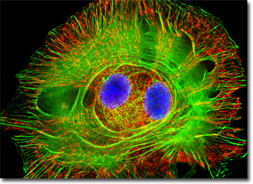
The African green monkey kidney cell that appears in the digital image above was resident in a CV-1 cell culture that was stained with MitoTracker Red CMXRos, DAPI, and Alexa Fluor 488 conjugated to phalloidin, which target the mitochondrial network, nuclear DNA, and filamentous actin, respectively. Images were recorded in grayscale with a QImaging Retiga Fast-EXi camera system coupled to an Olympus BX-51 microscope equipped with bandpass emission fluorescence filter optical blocks provided by Omega Optical. During the processing stage, individual image channels were pseudocolored with RGB values corresponding to each of the fluorophore emission spectral profiles.
Additional Fluorescence Images of African Green Monkey Kidney (CV-1) Cells
CV-1 Cells with Oregon Green 488, Alexa Fluor 568, and DAPI - A culture of CV-1 fibroblast cells was stained with wheat germ agglutinin conjugated to Oregon Green 488, Alexa Fluor 568 conjugated to phalloidin, and DAPI, which selectively bind to the intracellular Golgi network, cytoskeletal actin filaments, and nuclei, respectively. Note the presence of high signal levels, with sharp definition, from all three of the fluorophores employed to stain the specimen.
Actin Network in African Green Monkey Kidney Cells - Reviewed in this section is a culture of African green monkey kidney fibroblast cells that was stained with Alexa Fluor 488 conjugated to phalloidin, which binds to the intracellular filamentous actin network. The cells were subsequently stained with DAPI (targeting DNA in the cell nuclei) and the lectin, wheat germ agglutinin, conjugated to Texas Red (targeting glycoproteins in the Golgi complex).
African Green Monkey Kidney Fibroblast Cells with MitoTracker Red CMXRos, SYTOX Green, and Alexa Fluor 350 - The isolated cell that appears in this section was a resident in an adherent monolayer culture of CV-1 cells labeled with SYTOX Green to stain chromatin in the nuclei. In addition, the culture was treated with Alexa Fluor 350 conjugated to phalloidin and MitoTracker Red CMXRos, to target filamentous actin and mitochondrial network, respectively.
CV-1 Cellular Adherens Junction Cadherin with Cy3 - Normal (non-transformed) African green monkey kidney fibroblast cells (CV-1 line) were immunofluorescently labeled with primary anti-cadherin monoclonal antibodies (mouse) followed by goat anti-mouse Fab fragments conjugated to Cy3. In addition, the specimen was simultaneously stained for DNA with the ultraviolet-absorbing probe DAPI, and for the cytoskeletal filamentous actin network with Alexa Fluor 488 conjugated to phalloidin.
Microtubule and DNA Distribution in CV-1 Fibroblast Cells - A culture of African green monkey kidney fibroblasts was immunofluorescently labeled with primary anti-tubulin mouse monoclonal antibodies followed by goat anti-mouse Fab fragments conjugated to Rhodamine Red-X. In addition, the specimen was stained with DAPI (targeting DNA in the nucleus).
African Green Monkey Kidney Cells with MitoTracker Red CMXRos, Alexa Fluor 488, and DAPI - The isolated fibroblast that appears in this section was resident in a normal (non-transformed) African green monkey kidney cell culture labeled with MitoTracker Red CMXRos, Alexa Fluor 488 conjugated to phalloidin, and DAPI, targeting the mitochondrial network, F-actin, and nuclear DNA, respectively. Note the striking geometry of actin filaments, which demonstrate stress fibers, lamellipodia, filopodia, microspikes, and dorsal arcs.
CV-1 Cell Tubulin, Actin, and DNA with Alexa Fluor 568, Fluorescein, and DAPI - An adherent log phase monolayer CV-1 cell culture was immunofluorescently labeled with primary anti-bovine alpha-tubulin mouse monoclonal antibodies followed by goat anti-mouse Fab fragments conjugated to fluorescein. In addition, the cells were simultaneously stained for DNA with the ultraviolet-absorbing probe DAPI, and for the cytoskeletal filamentous actin network with Alexa Fluor 568 conjugated to phalloidin.
Focal Adhesion Sites in African Green Monkey Fibroblasts - A culture of African green monkey kidney cells was immunofluorescently labeled with primary anti-vinculin mouse monoclonal antibodies followed by goat anti-mouse Fab fragments conjugated to Cy3 (red emission). Vinculin is a protein associated with focal adhesion and adherens junctions, which are membrane-associated complexes that serve as nucleation sites for actin filaments and as crosslinkers between the external medium, plasma membrane, and actin cytoskeleton. The specimen was counterstained for F-actin with Alexa Fluor 488 conjugated to phalloidin, and for DNA with DAPI.
CV-1 Cytoskeletal Network Elements with Rhodamine Red-X, Alexa Fluor 488, and DAPI - In this section, the digital fluorescence microscopy image features CV-1 cells immunofluorescently labeled with primary anti-tubulin mouse monoclonal antibodies followed by goat anti-mouse Fab fragments conjugated to Rhodamine Red-X. In addition, the cells were stained with DAPI, which selectively binds to DNA in the cell nucleus, and Alexa Fluor 488 conjugated to phalloidin, which targets the filamentous actin network.
African Green Monkey Kidney Cells with MitoTracker Red CMXRos, Alexa Fluor 488, and DAPI - A log phase adherent culture of African green monkey kidney cells was stained with the classical cocktail: MitoTracker Red CMXRos, DAPI, and Alexa Fluor 488 conjugated to phalloidin, which target mitochondria, DNA in cell nuclei, and F-actin, respectively.
The Extensive Microtubule Network in CV-1 Cells - The microtubules present in a log phase culture of CV-1 cells were immunofluorescently labeled with primary anti-tubulin mouse monoclonal antibodies followed by goat anti-mouse Fab fragments conjugated to Rhodamine Red-X. In addition, the culture was simultaneously stained with DAPI, which selectively binds to DNA in the cell nucleus.
BACK TO THE CULTURED CELLS FLUORESCENCE GALLERY
BACK TO THE FLUORESCENCE GALLERY