Fluorescence Digital Image Gallery
Normal African Green Monkey Kidney Fibroblast Cells (CV-1)
|
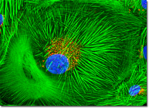
The primary cells of connective tissue are fibroblasts. Found throughout the body, connective tissue exhibits a smaller number of cells than most other tissue types and a significantly larger amount of intercellular substance. In addition to fibroblasts, macrophages, adipose cells, mast cells, lymphocytes, monocytes, eosinophils, and plasma cells are also often present in connective tissue. The connective tissue found in the cortex of the kidney, however, is believed to predominantly consist of fibroblasts and macrophages. Forming a capsule around the organ, this renal tissue is organized into a thin layer but is relatively dense and collagenous. The medullary interstitial connective tissue found in the kidney is significantly thicker than that of the cortex and vascular components and some apparatus of the uriniferous tubules are embedded in the material. Illustrated above is a culture of African green monkey kidney fibroblast cells that was stained with Alexa Fluor 488 conjugated to phalloidin, which binds to the intracellular filamentous actin network. The cells were subsequently stained with DAPI (targeting DNA in the cell nuclei) and the lectin, wheat germ agglutinin, conjugated to Texas Red (targeting glycoproteins in the Golgi complex). Images were recorded in grayscale with a QImaging Retiga Fast-EXi camera system coupled to an Olympus BX-51 microscope equipped with bandpass emission fluorescence filter optical blocks provided by Omega Optical. During the processing stage, individual image channels were pseudocolored with RGB values corresponding to each of the fluorophore emission spectral profiles. |
© 1995-2022 by Michael W. Davidson and The Florida State University. All Rights Reserved. No images, graphics, software, scripts, or applets may be reproduced or used in any manner without permission from the copyright holders. Use of this website means you agree to all of the Legal Terms and Conditions set forth by the owners.
This website is maintained by our
|