
Fluorescence Digital Image Gallery
Embryonic Swiss Mouse Fibroblast Cells (3T3 Line)
Established by George Todaro and Howard Green in 1962 from disaggregated Swiss mouse (Mus musculus) embryo tissue, the 3T3 cell line is a standard fibroblast cell line used in a wide spectrum of research and industrial biomedical applications. Variants of the initial cell line have been tested and found negative for ectromelia virus (mousepox), but most are susceptible to polyoma and simian virus 40 (SV40).
In addition, 3T3 cells are negative for reverse transcriptase, indicating the lack of integral retrovirus genomes. Within the cytoplasm, lysophosphatidylcholine (lyso-PC) induces AP-1 activity and c-jun N-terminal kinase activity (JNK1) by a protein kinase C-independent pathway. Contact inhibited, a confluent monolayer of 3T3 cells yields approximately 40,000 cells per square centimeter.
At the time of their establishment, 3T3 cells were different than most other cell lines in regard to the fact they did not induce tumors to develop when injected into murine species. However, 3T3 cells, it was quickly realized, were not normal cells either, since they are capable of growing indefinitely. In fact, the unusual behavior of the line enabled researchers to make a clear distinction for the first time between immortal cells and cells that have the ability to form tumors; previous to studies of the 3T3 line, it was believed that these characteristics were necessarily synonymous with one another. However, due to examination of 3T3 cells and subsequent research it has become widely accepted that for immortalization of cells to take place, telomere shortening, which can instigate chromosomal rearrangements, must be overcome, a process that is not necessarily related to a cell�s ability to undergo oncogenic transformation.
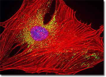
The isolated 3T3 cell presented in the digital image above was resident in an adherent culture stained for F-actin with Alexa Fluor 568 conjugated to phalloidin, and for DNA with 4',6-diamidino-2-phenylindole (DAPI). In addition, the culture was immunofluorescently labeled with Cy2 conjugated to antibodies that target peroxisomal membrane protein 70 (PMP 70), an abundant and integral membrane component of peroxisomes. Images were recorded in grayscale with a QImaging Retiga Fast-EXi camera system coupled to an Olympus BX-51 microscope equipped with bandpass emission fluorescence filter optical blocks provided by Omega Optical. During the processing stage, individual image channels were pseudocolored with RGB values corresponding to each of the fluorophore emission spectral profiles.
Additional Fluorescence Images of Embryonic Swiss Mouse Fibroblast (3T3) Cells
Swiss Mouse Embryo Fibroblast Cells with MitoTracker Red CMXRos, Alexa Fluor 488, and DAPI - A log phase culture of embryonic Swiss mouse fibroblast cells was stained with MitoTracker Red CMXRos, Alexa Fluor 488 conjugated to phalloidin, and DAPI, which target the intracellular mitochondrial network, cytoskeletal actin filaments, and nuclei, respectively. High signal levels from all three of the fluorophores employed to stain the culture are present.
Tubulin, Actin, and DNA Distribution in 3T3 Cells - A culture of 3T3 cells was immunofluorescently labeled with primary anti-tubulin mouse monoclonal antibodies followed by goat anti-mouse Fab fragments conjugated to the cyanine dye, Cy3. In addition, the cells were simultaneously probed for DNA with the ultraviolet-absorbing probe DAPI, and for the cytoskeletal filamentous actin network with Alexa Fluor 488 conjugated to phalloidin.
Visualizing Structural Features of the Golgi Complex and Nucleus in Swiss Mouse Embryo Cells - In order to examine structural features of the Golgi complex and nucleus at relatively high magnification, a log-phase culture of 3T3 cells was fixed, permeabilized, blocked with normal goat serum, and then treated with rabbit anti-giantin (Golgi protein) primary antibodies followed by goat anti-rabbit secondary antibodies (IgG) conjugated to Alexa Fluor 568. The nuclei were counterstained with Hoechst 33258.
Mouse Embryo Fibroblast Cells with Texas Red-X, Alexa Fluor 488, and DAPI - In this section, a Swiss mouse embryo fibroblast cell is presented that was resident in a culture labeled with the fluorophore Texas Red-X conjugated to wheat germ agglutinin, a fluorescent lectin that selectively binds to sialic acid residues. Wheat germ agglutinin conjugates are often used as probes for the Golgi network in mammalian cultures. The cells were also stained with Alexa Fluor 488 conjugated to phalloidin and DAPI, which target F-actin and DNA, respectively.
Labeling the Actin Cytoskeleton and Microtubule Network in 3T3 Swiss Mouse Embryo Cell Cultures - A monolayer culture of Swiss mouse embryo cells was immunofluorescently labeled with primary mouse anti-beta-tubulin antibodies, and then subsequently treated with a mixture of secondary antibodies conjugated to Alexa Fluor 568 in a mixture containing phalloidin conjugated to Alexa Fluor 350. The cell nuclei were counterstained with SYTOX Green.
Mouse Embryo Fibroblast Cells with Cy3, Alexa Fluor 488, and Hoechst 33342 - A culture of Swiss mouse embryo cells was immunofluorescently labeled with primary anti-vinculin mouse monoclonal antibodies followed by goat anti-mouse Fab fragments conjugated to Cy3 (yielding red emission). In addition, the specimen was simultaneously stained for DNA with the ultraviolet-absorbing probe Hoechst 33342 (blue emission), and for the cytoskeletal filamentous actin network with Alexa Fluor 488 (green emission) conjugated to phalloidin.
3T3 Cells with Alexa Fluor 568, Alexa Fluor 488, and DAPI - Alexa Fluor 568 conjugated to phalloidin was utilized to label a 3T3 cell culture for intracellular filamentous actin, while DAPI was used to target DNA in the cell nuclei. The cells were simultaneously stained with Alexa Fluor 488 conjugated to concanavalin A, a lectin displaying very high affinity for specific carbohydrate residues in glycoproteins, enzymes, and cell membranes.
Actin and Mitochondrial Networks in Embryonic Mouse Fibroblast Cell Cultures - The nuclei of embryonic Swiss mouse fibroblasts in culture were targeted with the nucleic acid probe DAPI, which has an excitation maximum at 358 nanometers and an emission maximum at 461 nanometers when bound to DNA in cell cultures and tissue sections. In addition, the cells were also stained with Alexa Fluor 488 conjugated to phalloidin (filamentous actin) and MitoTracker Red CMXRos (mitochondria).
Swiss Mouse Embryo 3T3 Cells with Alexa Fluor 568, Cy2, and Hoechst 33258 - A culture of 3T3 cells was labeled with Hoechst 33258, which selectively binds to DNA in the cell nucleus (blue emission), and Alexa Fluor 568 conjugated to phalloidin, which binds to the cytoskeletal filamentous actin network (red emission). The culture, a single resident cell of which appears in this section, was also probed immunofluorescently with primary anti-oxphos complex V inhibitor protein monoclonal antibodies followed by goat anti-mouse Fab fragments conjugated to Cy2 (green emission).
Swiss Mouse Embryo Fibroblasts with Rhodamine Red, Alexa Fluor 488, and Hoechst 33258 - Presented in this section is an adherent culture of Swiss mouse embryo fibroblasts that was immunofluorescently labeled with Rhodamine Red conjugated to antibodies directed against peroxisomal membrane protein 70 (PMP 70), an abundant and integral membrane component of peroxisomes. Alexa Fluor 488 conjugated to phalloidin and Hoechst 33258 were simultaneously used to counterstain the culture, targeting F-actin and DNA, respectively.
The Golgi Complex in 3T3 Cells - A log phase culture of 3T3 cells was immunofluorescently labeled with primary anti-human golgin-97 mouse monoclonal antibodies followed by goat anti-mouse Fab fragments conjugated to Alexa Fluor 488 in order to target the Golgi apparatus. In addition, the cells were labeled with Alexa Fluor 568 conjugated to phalloidin for filamentous actin and Hoechst 33258 for DNA in cell nuclei.
3T3 Swiss Mouse Fibroblasts with MitoTracker Red CMXRos, Alexa Fluor 488, and DAPI - The digital image of 3T3 cells displayed in this section clearly differentiates the mitochondria, filamentous actin networks, and nuclei present in the fibroblasts. The probes utilized to label the cells include MitoTracker Red CMXRos (red emission), Alexa Fluor 488 conjugated to phalloidin (green emission), and DAPI (blue emission).
Swiss Mouse Embryo Fibroblast Cells with Texas Red, Alexa Fluor 488, and DAPI - A single Swiss mouse embryo fibroblast cell that was part of an adherent culture stained with Texas Red conjugated to the lectin concanavalin A is presented in this section. Concanavalin A selectively binds to alpha-mannopyranosyl and alpha-glucopyranosyl residues in glycoproteins. Alexa Fluor 488 conjugated to phalloidin and DAPI were also used to label the culture, targeting filamentous actin and nuclear DNA, respectively.
Proximity of the Golgi Complex and Nucleus in 3T3 Monolayer Cell Cultures - The close proximity between the Golgi complex and nuclei in a culture of adherent albino Swiss mouse embryo cells was probed in a double immunofluorescence experiment with mouse anti-NPCP (nuclear pore complex protein) and rabbit anti-giantin primary antibodies. The antibody targets were visualized with goat secondary antibodies conjugated to Alexa Fluor 568 and Alexa Fluor 488, respectively, while the actin cytoskeletal framework was labeled with Alexa Fluor 350 conjugated to phalloidin.
Swiss Mouse Embryo Cellular Tubulin, Actin, and DNA - Alexa Fluor 568 conjugated to phalloidin, which targets cytoskeletal filamentous actin, and DAPI, which binds to DNA in cell nuclei, were used to probe a culture of Swiss mouse embryo fibroblast cells. In addition, the 3T3 cells were immunofluorescently labeled with primary anti-tubulin mouse monoclonal antibodies followed by goat anti-mouse Fab fragments conjugated to Cy2, targeting the microtubule network.
Adhesion Junctions in Swiss Mouse Embryo (3T3) Cells - An adherent culture of Swiss mouse embryo cells was immunofluorescently labeled with primary anti-vinculin mouse monoclonal antibodies followed by goat anti-mouse Fab heavy and light chain fragments conjugated to Cy3 (red emission). In addition, the specimen was simultaneously stained for DNA with the ultraviolet-absorbing probe Hoechst 33342, and for the cytoskeletal filamentous actin network with Alexa Fluor 488 conjugated to phalloidin.
Beta-Catenin Linking Proteins in 3T3 Cells - Log phase Swiss mouse embryo cells were immunofluorescently labeled with primary anti-beta-catenin rabbit monoclonal antibodies followed by goat anti-rabbit secondary antibodies conjugated to Rhodamine Red. In addition, nuclear histone proteins were targeted with anti-histone (pan) monoclonal antibodies, which were imaged with goat anti-mouse IgG (H + L) conjugated to Marina Blue (labeling the nucleus). The specimen was subsequently counterstained for the cytoskeletal filamentous actin network with Alexa Fluor 488 conjugated to phalloidin.
Targeting the Golgi Complex in 3T3 Swiss Mouse Embryo Cell Cultures with Immunofluorescence - A semi-confluent culture of 3T3 cells was fixed, permeabilized, and blocked with 10-percent normal goat serum in phosphate-buffered saline prior to immunofluorescent labeling with primary antibodies to giantin, a protein resident in the Golgi complex of mammalian cells. The culture was subsequently stained with a mixture of secondary antibodies conjugated to Alexa Fluor 488 in a mixture containing phalloidin conjugated to Alexa Fluor 568. The cell nuclei were counterstained with Hoechst 33342.
Targeting the Peroxisomal Membrane Proteins and Filamentous Actin Network in 3T3 Cells - Similar to one of the images linked above, this section reviews an adherent culture of Swiss mouse embryo fibroblasts that was immunofluorescently labeled with Rhodamine Red conjugated to antibodies directed against peroxisomal membrane protein 70 (PMP 70), an abundant and integral membrane component of peroxisomes. Alexa Fluor 488 conjugated to phalloidin and Hoechst 33258 were simultaneously used to counterstain the culture, targeting F-actin and DNA, respectively.
The Microtubule and Actin Cytoskeletal Networks in Swiss Mouse Embryo Cells - A monolayer culture of Swiss mouse embryo cells was immunofluorescently labeled with primary mouse anti-beta-tubulin antibodies, and then subsequently treated with a mixture of secondary antibodies conjugated to Alexa Fluor 568 in a mixture containing phalloidin conjugated to Alexa Fluor 350. The cell nuclei were counterstained with SYTOX Green.
Subcellular Localization of the Mitochondria in 3T3 Cells with DsRed Fluorescent Protein - Log phase 3T3 cells were transfected with a recombinant plasmid vector containing a chimeric fusion gene product of DsRed fluorescent protein and the mitochondrial targeting sequence from subunit VIII of human cytochrome C oxidase. Stable transfectants were fixed, permeabilized, and treated with phalloidin conjugated to Alexa Fluor 488 before being counterstained with DAPI.
Albino Swiss Mouse Embryo Cells with MitoTracker Red CMXRos, Alexa Fluor 488, and Hoechst 33342 - A culture of 3T3 mouse embryo cells was triple-labeled with the standard protocol: MitoTracker Red CMXRos to target the mitochondria, Alexa Fluor 488 conjugated to phalloidin for the filamentous actin network, and Hoechst 33342 to stain DNA in the cell nuclei.
Visualizing the Nuclear Pore Complex in Swiss Mouse Embryo (3T3) Cell Cultures - Using a monoclonal antibody directed against the nuclear pore complex proteins, an adherent culture of Swiss mouse embryo cells was fixed, permeabilized, and treated with primary mouse antibodies followed by goat anti-mouse secondary antibodies conjugated to Alexa Fluor 568 (red fluorescence). The cells were subsequently counterstained with Alexa Fluor 488 conjugated to phalloidin (filamentous actin; green fluorescence) and Hoechst 33342 (DNA in the nucleus; blue fluorescence).
Distribution of Histones, Peroxisomes, and Filamentous Actin in 3T3 Cell Cultures - In a double immunofluorescence labeling experiment, an adherent culture of Swiss mouse embryo cells was treated with a cocktail of mouse anti-histones (pan) and rabbit anti-PMP 70 (peroxisomal membrane protein) primary antibodies, followed by goat anti-mouse and anti-rabbit secondary antibodies conjugated to Alexa Fluor 568 and Alexa Fluor 488, respectively, to target the nuclear histone proteins and peroxisomes. The filamentous actin network was imaged with Alexa Fluor 350 conjugated to phalloidin.
BACK TO THE CULTURED CELLS FLUORESCENCE GALLERY
BACK TO THE FLUORESCENCE GALLERY