Fluorescence Digital Image Gallery
Embryonic Swiss Mouse Fibroblast Cells (3T3)
|
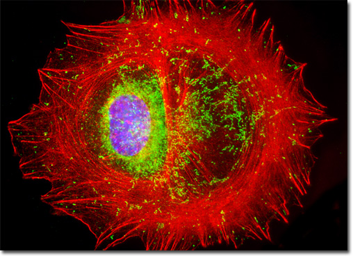
Established by George Todaro and Howard Green in 1962 from disaggregated Swiss mouse (Mus musculus) embryo tissue, the 3T3 cell line is a standard fibroblast cell line used in a wide spectrum of research and industrial biomedical applications. Variants of the initial cell line have been tested and found negative for ectromelia virus (mousepox), but most are susceptible to polyoma and simian virus 40 (SV40). In addition, 3T3 cells are negative for reverse transcriptase, indicating the lack of integral retrovirus genomes. Within the cytoplasm, lysophosphatidylcholine (lyso-PC) induces AP-1 activity and c-jun N-terminal kinase activity (JNK1) by a protein kinase C-independent pathway. Contact inhibited, a confluent monolayer of 3T3 cells yields approximately 40,000 cells per square centimeter. A culture of 3T3 cells was labeled with Hoechst 33258, which selectively binds to DNA in the cell nucleus (blue emission), and Alexa Fluor 568 conjugated to phalloidin, which binds to the cytoskeletal filamentous actin network (red emission). The culture, a single resident cell of which appears in the digital image above, was also probed immunofluorescently with primary anti-oxphos complex V inhibitor protein monoclonal antibodies followed by goat anti-mouse Fab fragments conjugated to Cy2 (green emission). Images were recorded in grayscale with a QImaging Retiga Fast-EXi camera system coupled to an Olympus BX-51 microscope equipped with bandpass emission fluorescence filter optical blocks provided by Omega Optical. During the processing stage, individual image channels were pseudocolored with RGB values corresponding to each of the fluorophore emission spectral profiles. |
© 1995-2025 by Michael W. Davidson and The Florida State University. All Rights Reserved. No images, graphics, software, scripts, or applets may be reproduced or used in any manner without permission from the copyright holders. Use of this website means you agree to all of the Legal Terms and Conditions set forth by the owners.
This website is maintained by our
|