
Bausch & Lomb Slit Ultramicroscope No. 22
In industry and materials science, the inspection of fine structure, such as the fibers in paper and textiles, the grain of wood, or reading finely divided scales, is dependent on the resolution of the optical instrument. The microscope illustrated below was fully described and accompanied by a photograph of an original Bausch & Lomb Slit Ultramicroscope No. 22 in the 1929 Bausch & Lomb catalog.
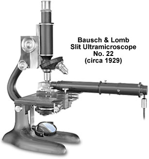
Based on the pioneering work by Dr. Heinrich Siedentopf in 1900, Bausch & Lomb design engineers devised the Slit Ultramicroscope No. 22 to go beyond the Abbe limit of optical resolution (approximately 0.20 micrometers) by illuminating "ultramicroscopic particles." During the late 1920s and early 1930s, the Bausch & Lomb Slit Ultramicroscope No. 22 reached a pinnacle for optical inspection microscopy. By coupling the Model FFS-6 monocular, compound microscope with a specially designed illumination system, the slit ultramicroscope is able to push beyond the Abbe limit of resolution for liquid, gaseous, or solid specimens. Specialized specimen holders are placed at the intersection of the perpendicular optical axes of the laboratory-grade microscope and the illumination system.
The ultramicroscope illumination system, mounted in a specialized 11-inch tube, consists of a light source, condensing system, and a very narrow slit. A 6-volt Mazda tungsten lamp, adjustable for centering and for proper focus, produces the light. The light source is imaged by the condensing system on the slit, which in turn is imaged by the objective in the object plane of the microscope. The slit width is altered by means of a small, knurled screw that is located at the midpoint of the illumination tube.
The slit ultramicroscope has two achromatic, water immersion objectives (44x, numerical aperture 1.00 and 26x, numerical aperture 0.50) mounted in a double, dust-proof, circular nosepiece. The RMS-threaded objectives are parfocal on their rotating nosepiece. Eyepieces with magnifications of either 5x or 10x, a 160-millimeter fixed-length body tube, an Abbe substage condenser (numerical aperture 1.20), and a 50-millimeter plano-concave mirror round out the optical train of the ultramicroscope.
BACK TO TWENTIETH CENTURY BAUSCH & LOMB MICROSCOPES
BACK TO TWENTIETH CENTURY MICROSCOPES