
Bausch & Lomb Chamot Chemical Microscope M
Solvent and reagent-proof throughout, the Bausch & Lomb Chamot Chemical Microscope M was designed based on specifications developed by Drs. Emil M. Chamot and C.W. Mason of the Laboratory of Chemical Microscopy at Cornell University in Ithaca, New York. The unique stand featured by the chemical microscope, which was novel for the period, allows more room for the manipulation of specimens.
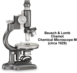
Other significant innovations for chemical microscopy include improved polarizing optics, and a much larger round, rotating stage with an optional mechanical stage. In addition, the microscope is equipped with a revolving nosepiece and a substage condenser assembly that is mounted on a rack and pinion mechanism. Achromatic objectives at 10x and 21x magnifications, each mounted on a dust-proof, near-parfocal, rotating nosepiece (as illustrated), are standard with the Chamot microscope. On the Model M-4, a triple nosepiece replaces the double circular nosepiece, and bears an additional 4x achromatic objective. There is also an optional centering nosepiece designed for the Chamot chemical microscope line. On each of the three chemical microscope models, which were manufactured during the late 1920s and early 1930s, the Huygenian eyepieces are fitted with cross hair reticles so that the planes of vibration of the polarizers and analyzers can be tracked.
All Bausch & Lomb Chamot chemical microscope M models feature oculars with magnifications of either 10x or 12.5x, while the M-4 is also equipped with eyepieces of 5x and 7.5x magnifications. The standard polarizer for parallel polarized light studies is a Nicol prism that is set in a revolving mount, while a rotatable Glan-Thompson prism, which slips over the eyepiece tube, performs as the analyzer. An optional F polarizer, consisting of a Nicol prism in a graduated, revolving mount, is engaged for convergent polarized light microscopy (primarily for describing interference figures or conoscopic images). The entire polarizer apparatus drops into the substage ring and is fixed perpendicular to the operator. The condenser and attached iris diaphragm, which are mounted on a pivoted arm of the F polarizer, can easily be swung out of the light pathway for parallel illumination. The substage ring also accommodates any choice from the complete line of Bausch & Lomb condensers. A standard 50-millimeter diameter plano-concave mirror completes the optical train for the Chamot chemical microscope.
Like many Bausch & Lomb monocular, compound microscopes, the Chamot chemical microscope M features coarse focusing that is accomplished with a rack and pinion mechanism. Large, knurled, brass pinion heads are positioned on either side of the body tube for easier and more comfortable focusing, even while viewing specimens through the eyepiece. The Bausch & Lomb patented fine-focus lever is graduated for vertical movements and for use in the determination of the refractive indexes of liquids. The circular stage is centerable utilizing two screws, and the circumference of the 102-millimeter diameter stage is graduated in single degrees, with every tenth degree numbered. An optional mechanical stage may be attached to the standard revolving stage. Alternatively, the standard stage may be removed for the substitution of the optional metallurgical auxiliary stage, a heating or cooling stage, or to facilitate rapid and thorough cleaning in case of chemical spills.
BACK TO TWENTIETH CENTURY BAUSCH & LOMB MICROSCOPES
BACK TO TWENTIETH CENTURY MICROSCOPES