
Chemical Crystal Movie Gallery
Niacinamide Time Lapse Sequences
Niacinamide, a more water-soluble form of vitamin B-3, may play a key role in fighting aging and repairing damaged joint cartilage, which is typical in arthritis. As the amide form of niacin, niacinamide may prevent, and in some cases even reverse, Type 1 (insulin-dependent) diabetes.
Known also as nicotinamide, niacinamide does not produce flushing of the skin, as does niacin when doses exceed 50 milligrams. Natural sources of niacinamide include beef liver, brewer's yeast, halibut, chicken, sunflower seeds, and peanuts. Synthesized niacinamide is marketed in the form of tablets, capsules, oral solutions, and as injectable formulations, and reportedly aids the release of energy from consumed foods and promotes DNA biosynthesis. In the digestion of fats, production of sugars, and in tissue respiration, the coenzymes NAD and NADP incorporate available niacinamide into their structures. However, while niacin also helps regulate cholesterol, niacinamide does not.
At higher concentrations than the 25 milligrams per 2 pounds of body weight prescribed for diabetes, niacinamide acts as a natural tranquilizer and binds onto the same brain receptor sites as synthesized pharmaceuticals such as Valium. Certain research suggests that niacinamide protects the liver against cirrhosis and other alcohol-abuse induced damage, and some trials of the chemical indicate that when it is ingested four times a day at 500-milligram doses, canker sores are inhibited and healed. Deficiencies in vitamin B-3 result in pellagra, headaches, depression, and other symptoms, and because cigarette smoking decreases vitamin B-3 absorption, smokers may require supplemental niacin. Overdoses of niacinamide cause vomiting and diarrhea and can result in high blood sugar, high uric acid, liver damage, and heart arrhythmia.
Niacinamide Time Lapse Sequence #1 - A time-lapse sequence of 18 images illustrating formation of spherulitic crystallites from a melt.
Niacinamide Time Lapse Sequence #2 - Spherulites of a variety of sizes crystallize and merge to fill the viewfield in this 19-image time-lapse sequence.
Niacinamide Time Lapse Sequence #3 - A spherulitic phase displaying a variety of textures forms from the melt in this sequence of 30 images.
Niacinamide Time Lapse Sequence #4 - This sequence of 17 time-lapse images illustrates the growth of crystalline spherulites that exhibit a change of texture as they reach larger sizes.
Niacinamide Time Lapse Sequence #5 - Various sizes of spherulites crystallize from the melt and merge into a confluent texture displaying blue to violet birefringence in a sequence of 17 images.
Niacinamide Time Lapse Sequence #6 - Two initial crystallization sites grow dramatically in size and are met by a growing field of much smaller spherulites in a time-lapse sequence of 26 images.
Niacinamide Time Lapse Sequence #7 - A 14-image time-lapse sequence illustrates crystal growth that begins with a field of small crystallites in the lower right-hand corner of the viewfield, followed by mixed-size spherulite formation throughout the remainder of the field.
Niacinamide Time Lapse Sequence #8 - Nucleation and growth of crystallites occurs more or less uniformly across the microscope viewfield in a time-lapse sequence of 12 images.
Niacinamide Time Lapse Sequence #9 - Spherulite formation occurs and advances from numerous locations across the viewfield in a time-lapse sequence of 15 images.
Niacinamide Time Lapse Sequence #10 - Various sizes of spherulitic crystallites grow throughout the microscope viewfield in a sequence of 9 images.
Niacinamide Time Lapse Sequence #11 - A time-lapse sequence of 26 images illustrates crystallization of a wide range of sizes of spherulites that merge into a field of varied texture exhibiting gradations of birefringence color.
Niacinamide Time Lapse Sequence #12 - A nearly uniform blue birefringence is displayed across the viewfield as crystallization occurs in a sequence of 10 time-lapse images.
Niacinamide Time Lapse Sequence #13 - A 13-image sequence of images illustrates crystallization of spherulites exhibiting blue to violet birefringence.
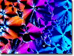
Niacinamide Time Lapse Sequence #14 - A kaleidoscopic display of birefringence colors occurs in this sequence of 26 time-lapse images illustrating the growth of spherulites.
Niacinamide Time Lapse Sequence #15 - Growth fronts of crystallization appear to advance toward each other from opposite sides of the viewfield in a time-lapse sequence of 27 images.
Niacinamide Time Lapse Sequence #16 - Portions of very large spherulitic crystals appear as fan shapes as they advance and merge with smaller crystallites in a time-lapse sequence of 24 images.
Contributing Authors
Omar Alvarado, Thomas J. Fellers and Michael W. Davidson - National High Magnetic Field Laboratory, 1800 East Paul Dirac Dr., The Florida State University, Tallahassee, Florida, 32310.
BACK TO THE CHEMICAL CRYSTAL MOVIE GALLERY
BACK TO THE DIGITAL IMAGE GALLERIES
Questions or comments? Send us an email.
© 1995-2022 by Michael W. Davidson and The Florida State University. All Rights Reserved. No images, graphics, software, scripts, or applets may be reproduced or used in any manner without permission from the copyright holders. Use of this website means you agree to all of the Legal Terms and Conditions set forth by the owners.
This website is maintained by our
Graphics & Web Programming Team
in collaboration with Optical Microscopy at the
National High Magnetic Field Laboratory.
Last Modification Friday, Nov 13, 2015 at 02:19 PM
Access Count Since September 17, 2002: 22275
Visit the website of our partner in introductory microscopy education:
|
|