
QX3 Stage And Stand
The stand of the Intel QX3 computer microscope is designed to hold the microscope body with a set of clamps in addition to supporting the stage with its focusing mechanism and the substage illuminator assembly. A cutaway drawing of the entire stand is illustrated in Figure 1. Electrical power for the substage illuminator is derived from a set of contacts located on the stand limb behind the body clamps.

A simple horseshoe stand design follows the basic theme used for a majority of the microscopes produced around the world during the past several hundred years on both toy and laboratory microscopes. The stand is made of an injection-molded high-impact polymer that is very lightweight and somewhat flexible. When the body is removed, the complete stand assembly weighs only about 330 grams (11.8 ounces), rendering it very susceptible vibrations and strong wind currents. Also, without any significant weight in the microscope base, the unit has a high center of gravity and is not difficult to tip over.
Another problem that we have discovered with this microscope is the lack of friction provided by the smooth plastic surface on the underside of the base. When placed on another smooth surface, such as a varnished table, the entire unit has a tendency to slide while the specimen is being loaded and unloaded or when the operator is too quick with the focus knobs. These problems can be remedied by adding weight to the base as we describe in the section on base modifications.

Focus is achieved with a rack and pinion mechanism that is composed around a set of nylon gears as illustrated above in Figure 2. The focusing knobs are stylishly fashioned from a combination of opaque and translucent blue plastic. They are attached to the pinion gear with a narrow stainless steel bar that traverses the center of the pinion gear and press-fits into a tubular hole in the focusing knobs.
Tension is maintained on the gearset by a spring and lever mounted on the inside microscope body limb with an open bearing that rides on the outer rod housing of the right-hand focus knob. This assembly adds stiffness to the up and down movement of the stage and prevents it from falling under the weight of heavy specimens. The spring tension should never be in need of adjustment, but it can be easily removed when the stand is disassembled. Bending the spring to a slightly larger angle will increase the tension, while reducing the spring angle will decrease tension--although this is not recommended.

Molded into the underside of the stage is the housing for a mixing chamber for the light emitted from the diascopic tungsten lamp. The mixing chamber (illustrated in Figure 3) is secured into this housing using two Phillips-head screws. A high-gloss white paint is used to coat the inside of the mixing chamber to ensure proper mixing of light emitted by the lamp.
Light mixed in the chamber exits through the plastic diffusion screen, which is positioned to cover a 30 millimeter (one and one-eight inch) opening in the stage. This light provides illumination for viewing specimens in transmitted light mode.

Direct current necessary to power the tungsten lamp is derived through a set of electrical contacts (illustrated in Figure 4) that are mounted in the limb of the microscope stand. This power originates at the USB connector on the printed circuit board in the microscope body and is transferred to the stand through another set of contacts that that are housed on the body. When the body is inserted into the stand, the two sets of electrical contacts are positioned to come together to provide continuity to close the circuit. The electricity travels to the lamp through a set of wires hidden internally (Figure 4) in the stand.
The tungsten lamp in the mixing chamber has a finite lifespan and must be replaced from time to time. Mattel provides a customer service telephone number in the literature accompanying the Intel QX3 microscope, which lists the spare parts center from which new lamps can be ordered. To replace a defective lamp, unscrew the mixing chamber from the underside of the stage to expose the lamp housing. The lamp is fitted to the housing with two thin wire contacts that allow the lamp to be slipped into the housing. Replace the lamp and reattach the mixing chamber to the stage.
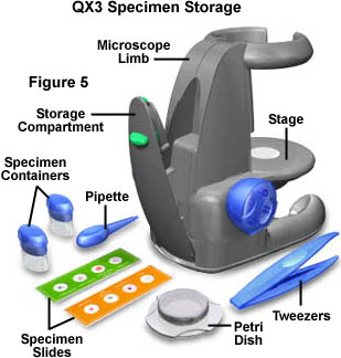
The Intel QX3 microscope has a variety of accessories to assist students in their microscopy investigations (as illustrated in Figure 5). These include a pipette (eyedropper), a pair of tweezers, two specimen containers, two covered Petri dishes, and a pair of pre-prepared microscope slides with four specimens each. The microscope slides are ready for observation as soon as the microscope is unpacked and the software installed. Specimens on the slides are a wool sample, Drosophila (fruit fly), shrimp eggs, sponge, spirogyra, fern spores, handmade paper, and dog hair. These specimens represent a great start in the search for objects to image with the microscope.
The specimen containers (Intel calls them sample jars) are great for collecting live insects, pond water samples, hairs, dirt, and other specimens that are candidates for microscopy. Pond water and crawling insects can be placed in the Petri dishes for observation in the microscope. The kit also includes a pair of tweezers for collecting specimens that children would not want to touch with their fingers, such as bugs, worms, and other small organisms. The pipette (eyedropper) is useful for transferring aliquots of pond water from the specimen containers to the Petri dishes.
The accessories will also help children understand that specimen collection and preparation are of paramount importance in microscopy. A handy storage compartment (Figure 5) is included in the microscope stand for storage of these accessories so they won't get lost.
In conclusion, the stand for the Intel Play QX3 computer microscope is a multi-functional unit that provides transmitted light illumination and a movable stage with which to focus the specimen. Modifications to the stand can greatly improve microscope performance and are addressed in a separate section entitled base modifications elsewhere in the Science, Optics & You website.
BACK TO INTEL PLAY QX3 MICROSCOPE ANATOMY
Questions or comments? Send us an email.
© 1995-2025 by Michael W. Davidson and The Florida State University. All Rights Reserved. No images, graphics, software, scripts, or applets may be reproduced or used in any manner without permission from the copyright holders. Use of this website means you agree to all of the Legal Terms and Conditions set forth by the owners.
This website is maintained by our
Graphics & Web Programming Team
in collaboration with Optical Microscopy at the
National High Magnetic Field Laboratory.
The QX3 microscope design is copyrighted © 2025 by Mattel, Inc. Intel® Play™ is a registered trademark of the Intel Corporation. These companies reserve all of their rights and privileges under copyright law. The material contained in this website is solely the opinion of the authors and is intended for eduational use only.
Last Modification Friday, Nov 13, 2015 at 01:18 PM
Access Count Since December 24, 1999: 39390
Visit the official Intel Play website:
![]()