
Advanced Condenser Systems: Abbe Condensers
Transmitted Darkfield Illumination
Digital Image Gallery
Darkfield microscopy relies on oblique illumination from the substage condenser to provide contrast to the specimen, which appears bright on a dark background. A central opaque stop is added to the light path beneath the condenser aperture and serves to block out the central illuminating rays from the light source.
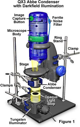
The remaining light rays strike the specimen at oblique angles from all azimuths, producing beautiful digital images of insects, fibers, aquatic organisms and other specimens that are difficult to image with brightfield illumination. A configuration utilizing the QX3 microscope body with an aftermarket Abbe condenser with a darkfield stop is illustrated in Figure 1. The microscope body and condenser are secured into place axially using ring stands and clamps. Beneath the condenser is an opaque darkfield stop of variable diameter, which is also secured to a ring stand with a clamp. Illumination is provided by a 120-volt 20-watt tungsten lamphouse. The illuminator has a removable color conversion filter that adjusts the 2900 K tungsten light to daylight illumination (5500 K). We have investigated optical microscopy in darkfield mode using the configuration presented in Figure 1 and have assembled a gallery of digital images created with the QX3 body coupled to an Abbe darkfield condenser. Use the links below to navigate to individual photomicrographs of specimens photographed with the QX3 computer microscope equipped for advanced darkfield illumination.
Adult Flea - Highlighted areas surrounding the head of an adult flea produce superior contrast and enhanced specimen detail when imaged using this advanced technique.
Butterfly Wings - These beautiful structures are composed of miniature scales that interact with light to cause interference, leading to the wide spectrum of colors observed in butterfly wings.
Centipede Head - Poison claws greet our visitors to this image, which was mounted as a whole specimen and captured with darkfield illumination.
Cretaceous Dinosaur Fossil - An unusual photomicrograph of a mineralized dinosaur bone fossil showing jagged teeth-shaped structures.
Flea Whole Mount - Fine details become visible when the QX3 microscope is used to image an entire flea in darkfield illumination.
House Fly Head - Get face-to-face with one of your favorite creatures. Although more abundant in the warm spring and hot summer weather, house flies may exist year-round in temperate climates, where their life cycles occur every eight days.
Human Joint Tissue - In vertebrates (including humans), joints are structures that separate two or more adjacent elements of the skeleton, allowing them to move. Darkfield microscopy is often useful for examining stained thin sections of tissue samples, and these images of tissue from a human joint show how a substage condenser can markedly improve QX3 digital images using darkfield illumination.
Lily Flower - The lily flower figures prominently as a symbol in mythology, folklore, and contemporary culture. A sliced section from this beautiful flower holds equal beauty under the microscope.
Tilia (Basswood) Stem - Stained thin sections from this ornamental hardwood tree produce beautiful images under darkfield illumination.
BACK TO THE INTEL PLAY ADVANCED DIGITAL IMAGE GALLERY
Questions or comments? Send us an email.
© 1995-2025 by Michael W. Davidson and The Florida State University. All Rights Reserved. No images, graphics, software, scripts, or applets may be reproduced or used in any manner without permission from the copyright holders. Use of this website means you agree to all of the Legal Terms and Conditions set forth by the owners.
This website is maintained by our
Graphics & Web Programming Team
in collaboration with Optical Microscopy at the
National High Magnetic Field Laboratory.
The QX3 microscope design is copyrighted © 2025 by Mattel, Inc. Intel® Play™ is a registered trademark of the Intel Corporation. These companies reserve all of their rights and privileges under copyright law. The material contained in this website is solely the opinion of the authors and is intended for eduational use only.
Last Modification Friday, Nov 13, 2015 at 02:19 PM
Access Count Since April 25, 2000: 24385
Visit the official Intel Play website:
![]()