Fluorescence Digital Image Gallery
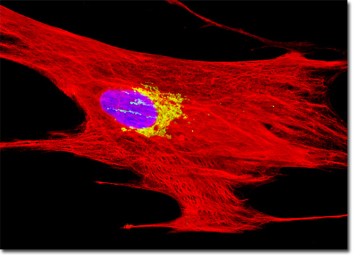
Horse Dermal Fibroblast Cells (NBL-6)
|
The intermediate filament is one of the three major types of filaments that comprise the cytoskeleton. Not all cells require the presence of the filaments, however, and they are notably absent from certain cell types, such as the oligodendrocytes present in the central nervous system of vertebrates. Intermediate filaments are most common and numerous in the cytoplasmic regions of cells that are frequently exposed to mechanical stress. Indeed, the primary role of the filaments appears to be to enhance cellular strength and durability, essentially maintaining the integrity of the cell. Several different types of intermediate filaments have been identified, all of which characteristically range in diameter from 8 to 10 nanometers. When the filaments occur in cells of mesenchymal origin, they are often composed of the polypeptide vimentin. The NBL-6 dermal fibroblast cells illustrated in the digital image above were fixed with paraformaldehyde, permeabilized, and treated with a mixture of rabbit (anti-giantin) and mouse (anti-vimentin; pan) primary antibodies, followed by secondary antibodies conjugated to Oregon Green 488 and Texas Red, respectively. Cell nuclei were counterstained with the DNA-selective bisbenzimide dye, Hoechst 33342. Images were recorded in grayscale with a QImaging Retiga Fast-EXi camera system coupled to an Olympus BX-51 microscope equipped with bandpass emission fluorescence filter optical blocks provided by Omega Optical. During the processing stage, individual image channels were pseudocolored with RGB values corresponding to each of the fluorophore emission spectral profiles. |
© 1995-2025 by Michael W. Davidson and The Florida State University. All Rights Reserved. No images, graphics, software, scripts, or applets may be reproduced or used in any manner without permission from the copyright holders. Use of this website means you agree to all of the Legal Terms and Conditions set forth by the owners.
This website is maintained by our
|