Fluorescence Digital Image Gallery
Horse Dermal Fibroblast Cells (NBL-6)
|
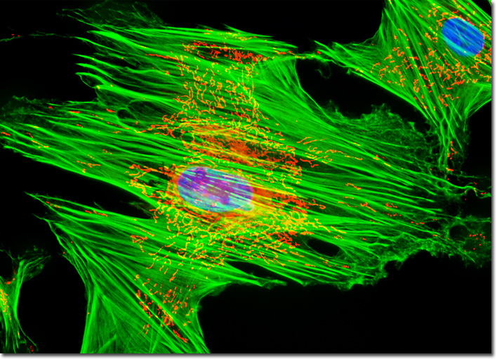
Each mitochondrion resident in a cell possesses two specialized membranes that function in different manners while segmenting the organelle into two separate compartments, namely an internal matrix and a narrow intermembrane space. The outer membrane of a mitochondrion acts a sieve due to the presence of numerous copies of the channel-forming protein porin in the lipid bilayer and is impenetrable to molecules greater than 5000 Daltons in size. The molecules that do pass through the outer membrane are usually impeded by the more-selective inner membrane. The inner membrane of the energy-generating organelle is typically highly convoluted and possesses a large amount of the phospholipid cardiolipin, which renders this lipid bilayer particularly difficult for ions to penetrate. In addition, the inner membrane contains proteins that carry out oxidation reactions, generate adenosine triphosphate (ATP) in the internal matrix, and enable metabolites to move into and out of the matrix. The mitochondrial network was targeted in the culture of NBL-6 fibroblasts featured in the digital image above with the X-rosamine derivative, MitoTracker Red CMXRos. The culture was simultaneously labeled for the cytoskeletal filamentous actin network and nuclear DNA with Alexa Fluor 488 conjugated to phalloidin and Hoechst 33258, respectively. Images were recorded in grayscale with a QImaging Retiga Fast-EXi camera system coupled to an Olympus BX-51 microscope equipped with bandpass emission fluorescence filter optical blocks provided by Omega Optical. During the processing stage, individual image channels were pseudocolored with RGB values corresponding to each of the fluorophore emission spectral profiles. |
© 1995-2025 by Michael W. Davidson and The Florida State University. All Rights Reserved. No images, graphics, software, scripts, or applets may be reproduced or used in any manner without permission from the copyright holders. Use of this website means you agree to all of the Legal Terms and Conditions set forth by the owners.
This website is maintained by our
|