Fluorescence Digital Image Gallery
Horse Dermal Fibroblast Cells (NBL-6)
|
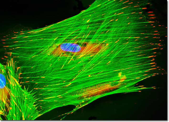
Vinculin is a ubiquitous cytoskeletal protein associated with cell adhesion and cell migration. A recent study carried out by a research group at St. Jude Children’s Research Hospital seems to indicate that the ability of vinculin to undergo precise changes in its shape may be central to understanding how the protein carries out such various roles. Such an understanding could have a tremendous impact on the medical world because the protein, which facilitates the movement of cells during embryonic development and for wound healing, also apparently plays a key role in the metastasis of cancers. According to the findings of the St. Jude study, vinculin alters its shape by moving the individual helical cylinder-like structures comprising its head in a process dubbed helical bundle conversion, which is sparked by the binding of one of two different proteins to the region. Talin apparently incites a shape change that is necessary for vinculin’s ability to anchor cells to the extracellular matrix, whereas the shape alteration caused by the presence of alpha-actinin facilitates the protein's ability to stabilize cadherin and, consequently, accumulate to form tissues and organs. The culture of horse dermal fibroblast cells presented in the digital image above was immunofluorescently labeled with anti-vinculin mouse monoclonal primary antibodies followed by goat anti-mouse Fab fragments conjugated to Alexa Fluor 568 (yielding red emission). In addition, the specimen was simultaneously stained for DNA with Hoechst 33342 (blue emission), and for the cytoskeletal filamentous actin network with Alexa Fluor 488 (green emission) conjugated to phalloidin. Images were recorded in grayscale with a QImaging Retiga Fast-EXi camera system coupled to an Olympus BX-51 microscope equipped with bandpass emission fluorescence filter optical blocks provided by Omega Optical. During the processing stage, individual image channels were pseudocolored with RGB values corresponding to each of the fluorophore emission spectral profiles. |
© 1995-2025 by Michael W. Davidson and The Florida State University. All Rights Reserved. No images, graphics, software, scripts, or applets may be reproduced or used in any manner without permission from the copyright holders. Use of this website means you agree to all of the Legal Terms and Conditions set forth by the owners.
This website is maintained by our
|