Fluorescence Digital Image Gallery
Madin-Darby Ovine Kidney Epithelial Cells (MDOK)
|
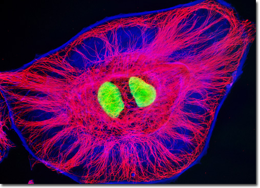
Although the cytoskeleton provides a certain amount of physical stability to the cell, it is a dynamic network. By adapting itself according to cellular needs, the cytoskeleton is able to form a remarkable array of structures from only three fundamental types of filaments. For example, the long, hollow, tube-like cytoskeletal filaments called microtubules can rearrange themselves rapidly from their typical cytoplasmic arrangement around the centrosome into a bipolar mitotic spindle when necessary for division of the cell. The microtubular elements of the cytoskeleton can also fashion whip-like flagella and cilia, while the relatively flexible actin filaments (also called microfilaments) are capable of forming into structures such as filopodia and lamellipodia. The third basic type of cytoskeletal filament, which is known as the intermediate filament due to the fact that its typical 8- to 12-nanometer diameter falls between that of microtubules (25 nanometers) and actin filaments (5 to 9 nanometers), lines the inside of the envelope surrounding the nucleus and can twist itself to increase its strength in order to maintain the integrity of a sheet of cells as well as facilitate the extension of long axons from neurons. A comparison between the microtubule and actin networks in MDOK cells (presented above) was conducted by treating a fixed and permeabilized adherent culture with mouse anti-tubulin primary antibodies, followed by a cocktail of goat anti-mouse secondary antibodies conjugated to Alexa Fluor 568 along with phalloidin conjugated to Alexa Fluor 350 (targeting the microtubule and filamentous actin networks, respectively). Nuclei were counterstained with SYTOX Green. Images were recorded in grayscale with a QImaging Retiga Fast-EXi camera system coupled to an Olympus BX-51 microscope equipped with bandpass emission fluorescence filter optical blocks provided by Omega Optical. During the processing stage, individual image channels were pseudocolored with RGB values corresponding to each of the fluorophore emission spectral profiles. |
© 1995-2025 by Michael W. Davidson and The Florida State University. All Rights Reserved. No images, graphics, software, scripts, or applets may be reproduced or used in any manner without permission from the copyright holders. Use of this website means you agree to all of the Legal Terms and Conditions set forth by the owners.
This website is maintained by our
|