Fluorescence Digital Image Gallery
Madin-Darby Ovine Kidney Epithelial Cells (MDOK)
|
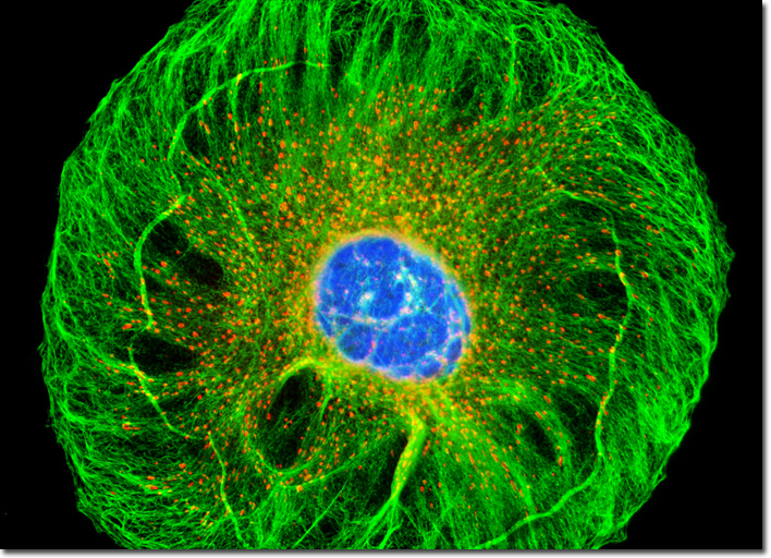
The intermediate filament is one of the three major filament families that comprise the cytoskeleton in vertebrate cells. The name of the filament refers to its diameter size (8 to 12 nanometers), which is intermediary between the diameters of the other two cytoskeletal filament varieties (actin filaments and microtubules). Intermediate filaments are designed to withstand significant amounts of tension and are less likely to disassemble and reassemble in the cell than other cytoskeletal filaments. The filaments are, therefore, relatively permanent cell structures and tend to be most prominent in cells exposed to significant physical stress. Indeed, when cell cultures are exposed to certain chemical treatments, microtubules and actin filaments disappear, but many networks of intermediate filaments remain structurally unchanged. Thus, intermediate filaments appear to be particularly vital in maintaining cell shape. The cytokeratin intermediate filament network in a culture of MDOK cells, illustrated above, was visualized by fixing an adherent culture in methanol, followed by treatment with mouse anti-cytokeratin (pan) monoclonal antibodies and goat anti-mouse Fab fragments conjugated to Cy2. Prior to fixation, the cells were exposed to MitoTracker Red CMXRos for one hour, and the culture was subsequently counterstained with Hoechst 33258 to image DNA in the nuclei. Images were recorded in grayscale with a QImaging Retiga Fast-EXi camera system coupled to an Olympus BX-51 microscope equipped with bandpass emission fluorescence filter optical blocks provided by Omega Optical. During the processing stage, individual image channels were pseudocolored with RGB values corresponding to each of the fluorophore emission spectral profiles. |
© 1995-2025 by Michael W. Davidson and The Florida State University. All Rights Reserved. No images, graphics, software, scripts, or applets may be reproduced or used in any manner without permission from the copyright holders. Use of this website means you agree to all of the Legal Terms and Conditions set forth by the owners.
This website is maintained by our
|