Fluorescence Digital Image Gallery
Madin-Darby Ovine Kidney Epithelial Cells (MDOK)
|
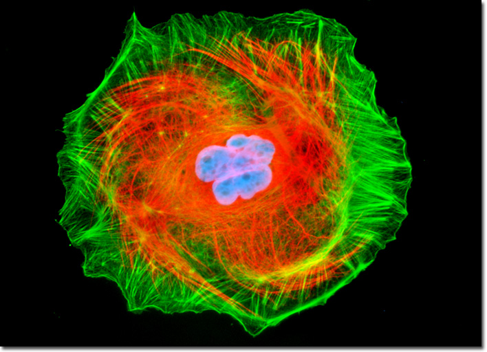
Vimentin is an intermediate filament protein (58 kiloDaltons) that is generally found in a variety of cells of mesenchymal origin and is developmentally regulated. Unlike most other proteins that comprise intermediate filaments, vimentin is expressed during the initial stages of cellular development, but is typically substituted with other tissue-specific intermediate filament proteins later in the developmental process. Vimentin has proven useful in the differential detection of undifferentiated neoplasms and is thought be involved in the communication and transport between the surface of a cell and its nucleus. An entire family of intermediate filaments, usually termed vimentin-like or, more simply, vimentin filaments, are named after the polypeptide. Vimentin-like intermediate filaments may also be composed of other proteins, however, such as desmin, peripherin, and glial fibrillary acidic protein (GFAP), which are closely homologous to vimentin. The log phase adherent culture of MDOK cells illustrated above was fixed and permeabilized with a cocktail of paraformaldehyde, Triton-X100, and glutaraldehyde before being blocked with 10-percent normal goat serum. Subsequent treatment with mouse anti-vimentin monoclonal primary antibodies was followed by goat anti-mouse secondary antibodies (IgG) conjugated to Texas Red-X. In addition, the filamentous actin was labeled with Oregon Green 488 conjugated to phalloidin and the nuclei counterstained with Hoechst 33258. Images were recorded in grayscale with a QImaging Retiga Fast-EXi camera system coupled to an Olympus BX-51 microscope equipped with bandpass emission fluorescence filter optical blocks provided by Omega Optical. During the processing stage, individual image channels were pseudocolored with RGB values corresponding to each of the fluorophore emission spectral profiles. |
© 1995-2025 by Michael W. Davidson and The Florida State University. All Rights Reserved. No images, graphics, software, scripts, or applets may be reproduced or used in any manner without permission from the copyright holders. Use of this website means you agree to all of the Legal Terms and Conditions set forth by the owners.
This website is maintained by our
|