Fluorescence Digital Image Gallery
Madin-Darby Canine Kidney Epithelial Cells (MDCK)
|
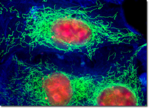
Plasmid pEYFP-Mitochondria vector gene product expression in various cell types occurs due to the efficient intracellular translation of a fusion nucleotide sequence combining the enhanced yellow fluorescent protein domain with the mitochondria targeting sequence from subunit VIII of human cytochrome C oxidase. The addition of simian virus 40 (SV40) polyadenylation signals, which are inserted downstream from the chimeric EYFP-Mitochondria fusion sequence, assures proper processing of the transcribed messenger RNA 3' terminus. The fluorescence excitation maximum of EYFP is 513 nanometers and the corresponding emission maximum occurs at 527 nanometers, with a relatively high (approximately 0.60) fluorescence quantum yield. In addition to the four chromophore mutations that shift the fluorescence emission maximum, the nucleotide coding sequence of the EYFP gene contains over 190 silent base alterations, which correspond to human codon-usage preferences that increase translational efficiency. The culture of Madin-Darby canine kidney epithelial cells appearing in the digital image above was transfected with a pEYFP-Mitochondria chimeric plasmid subcellular localization vector. The cells were also stained with SYTOX Orange and Alexa Fluor 350 conjugated to phalloidin, targeting DNA and the cytoskeletal filamentous actin network, respectively. Images were recorded in grayscale with a QImaging Retiga Fast-EXi camera system coupled to an Olympus BX-51 microscope equipped with bandpass emission fluorescence filter optical blocks provided by Omega Optical. During the processing stage, individual image channels were pseudocolored with RGB values corresponding to each of the fluorophore emission spectral profiles. |
© 1995-2025 by Michael W. Davidson and The Florida State University. All Rights Reserved. No images, graphics, software, scripts, or applets may be reproduced or used in any manner without permission from the copyright holders. Use of this website means you agree to all of the Legal Terms and Conditions set forth by the owners.
This website is maintained by our
|