Fluorescence Digital Image Gallery
Tahr Ovary Epithelial Cells (HJ1.Ov)
|
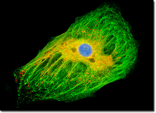
Unlike tubulin and actin, which are high conserved proteins that comprise microtubules and microfilaments (also known as actin filaments), respectively, the proteins that form intermediate filaments are very diverse. Indeed, the intermediate filament family of cytokeratins contains more than 20 different varieties of proteins that are found in human epithelial cells alone. Several different kinds of keratins may occur in the same epithelial cell, forming a continuous network through copolymerization. Despite their variety, all keratin filaments are comprised of tetramers formed from heterodimers of acidic type I keratin chains and neutral/basic type II keratin chains. Keratin filaments that are networked together are very stable, numerous disulfide bonds forming between the cross-linked elements. The formation of these strong bonds, which render keratin filaments insoluble in hot or cold water and enable them to outlive the cells in which they are housed to form hair, nails, and similar materials, is attributable to the large amount of the amino acid cystine contained in the proteins. The adherent log phase culture of tahr ovary cells illustrated above was treated for one hour with MitoTracker Red CMXRos in order to label the mitochondrial network, and the fixed cells were then incubated with mouse anti-cytokeratin primary antibodies followed by goat anti-mouse secondary antibodies (IgG) conjugated to Alexa Fluor 488. The nuclei were counterstained with Hoechst 33258. Images were recorded in grayscale with a Hamamatsu Orca-AG camera system coupled to an Olympus BX-51 microscope equipped with bandpass emission fluorescence filter optical blocks provided by Omega Optical. During the processing stage, individual image channels were pseudocolored with RGB values corresponding to each of the fluorophore emission spectral profiles. |
© 1995-2025 by Michael W. Davidson and The Florida State University. All Rights Reserved. No images, graphics, software, scripts, or applets may be reproduced or used in any manner without permission from the copyright holders. Use of this website means you agree to all of the Legal Terms and Conditions set forth by the owners.
This website is maintained by our
|