Fluorescence Digital Image Gallery
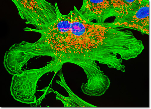
Tahr Ovary Epithelial Cells (HJ1.Ov)
|
As can be seen in the digital image featured above, mitochondria occupy a significant percentage of the cytoplasmic volume of eukaryotic cells. The important organelles, which frequently change shape and move about the cell, are considered to have been indespensible for the evolution of complex animals due to their key role in energy production. Indeed, if animal cells did not contain mitochondria, they would be required to solely depend upon anaerobic glycolysis for all ATP generation, a process that only releases a minute fraction of the potentially obtainable total free energy from glucose. By completing the metabolism of the sugar, mitochondria enable the generation of 15 times more ATP than simple anaerobic glycolysis. The adherent monolayer HJ1.Ov cell culture presented in the digital image above was labeled for the cytoskeletal filamentous actin and intracellular mitochondrial networks with Alexa Fluor 488 conjugated to phalloidin and MitoTracker Red CMXRos, respectively. Nuclei present in the epithelial cells were counterstained with the DNA-selective bisbenzimide dye, Hoechst 33258. Images were recorded in grayscale with a QImaging Retiga Fast-EXi camera system coupled to an Olympus BX-51 microscope equipped with bandpass emission fluorescence filter optical blocks provided by Omega Optical. During the processing stage, individual image channels were pseudocolored with RGB values corresponding to each of the fluorophore emission spectral profiles. |
© 1995-2025 by Michael W. Davidson and The Florida State University. All Rights Reserved. No images, graphics, software, scripts, or applets may be reproduced or used in any manner without permission from the copyright holders. Use of this website means you agree to all of the Legal Terms and Conditions set forth by the owners.
This website is maintained by our
|