Fluorescence Digital Image Gallery
Tahr Ovary Epithelial Cells (HJ1.Ov)
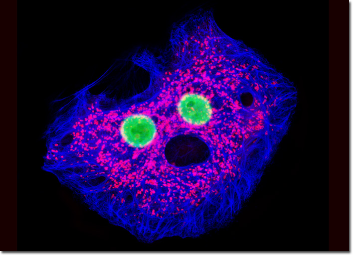
|
In recent years, keratins have been of significant interest to the medical community. The discovery that the diversity of the proteins can be utilized to provide additional information about the origination of carcinomas has made them popular clinical tools in cancer diagnosis. Also, genetic mutations in keratins have been identified as the cause of a variety of human disorders. The disease known as epidermolysis bullosa simplex, for example, is the result of a mutant keratin gene that causes the skin to be extremely fragile and to blister severely and recurrently. The blistering associated with this inherited disorder occurs due to the attempts of defective keratins to assemble with normal keratins in the epidermal basal cell layer, effectively disrupting the network of keratin filaments to the point of rupture. After treatment for one hour with MitoTracker Red CMXRos in serum-based growth medium, the adherent culture of HJ1.Ov cells presented above was fixed in 50:50 methanol:acetone, permeabilized, blocked, and treated with mouse anti-cytokeratin (pan) monoclonal antibodies. The intermediate filaments were visualized with goat anti-mouse secondary antibodies (IgG) conjugated to Marina Blue, and the nuclei were counterstained with SYTOX Green. Images were recorded in grayscale with a QImaging Retiga Fast-EXi camera system coupled to an Olympus BX-51 microscope equipped with bandpass emission fluorescence filter optical blocks provided by Omega Optical. During the processing stage, individual image channels were pseudocolored with RGB values corresponding to each of the fluorophore emission spectral profiles. |
© 1995-2025 by Michael W. Davidson and The Florida State University. All Rights Reserved. No images, graphics, software, scripts, or applets may be reproduced or used in any manner without permission from the copyright holders. Use of this website means you agree to all of the Legal Terms and Conditions set forth by the owners.
This website is maintained by our
|